Human Kidney Structure and Function — Anatomy, Filtration Process, and the Body’s Internal Regulation System
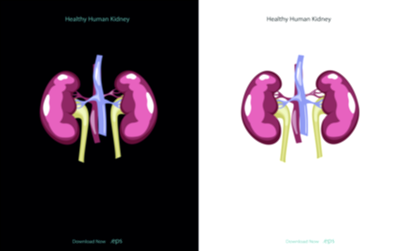
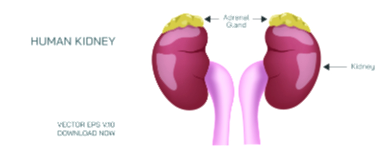
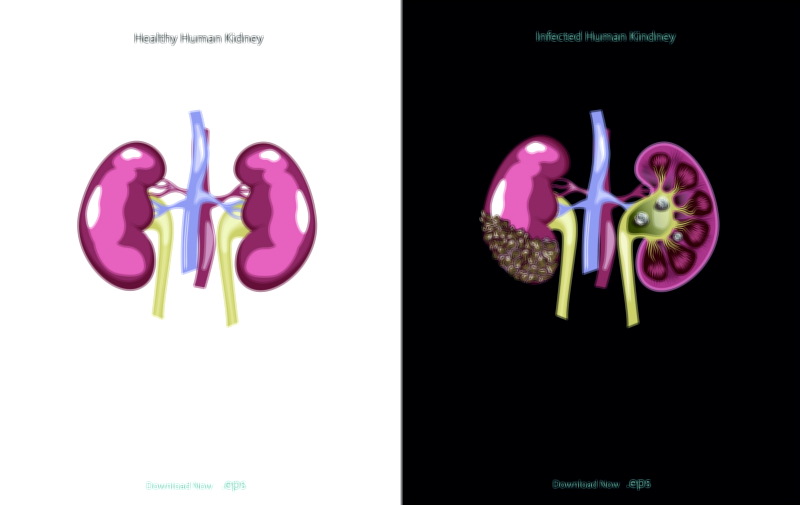
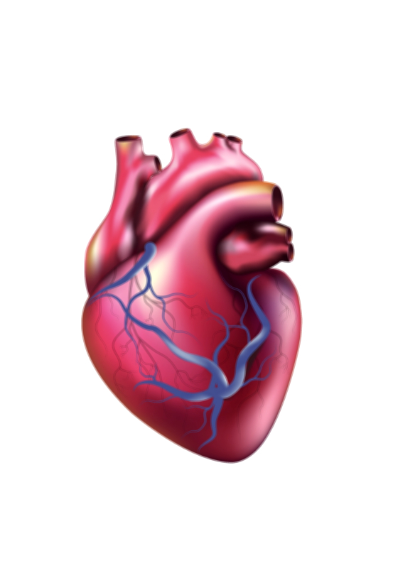
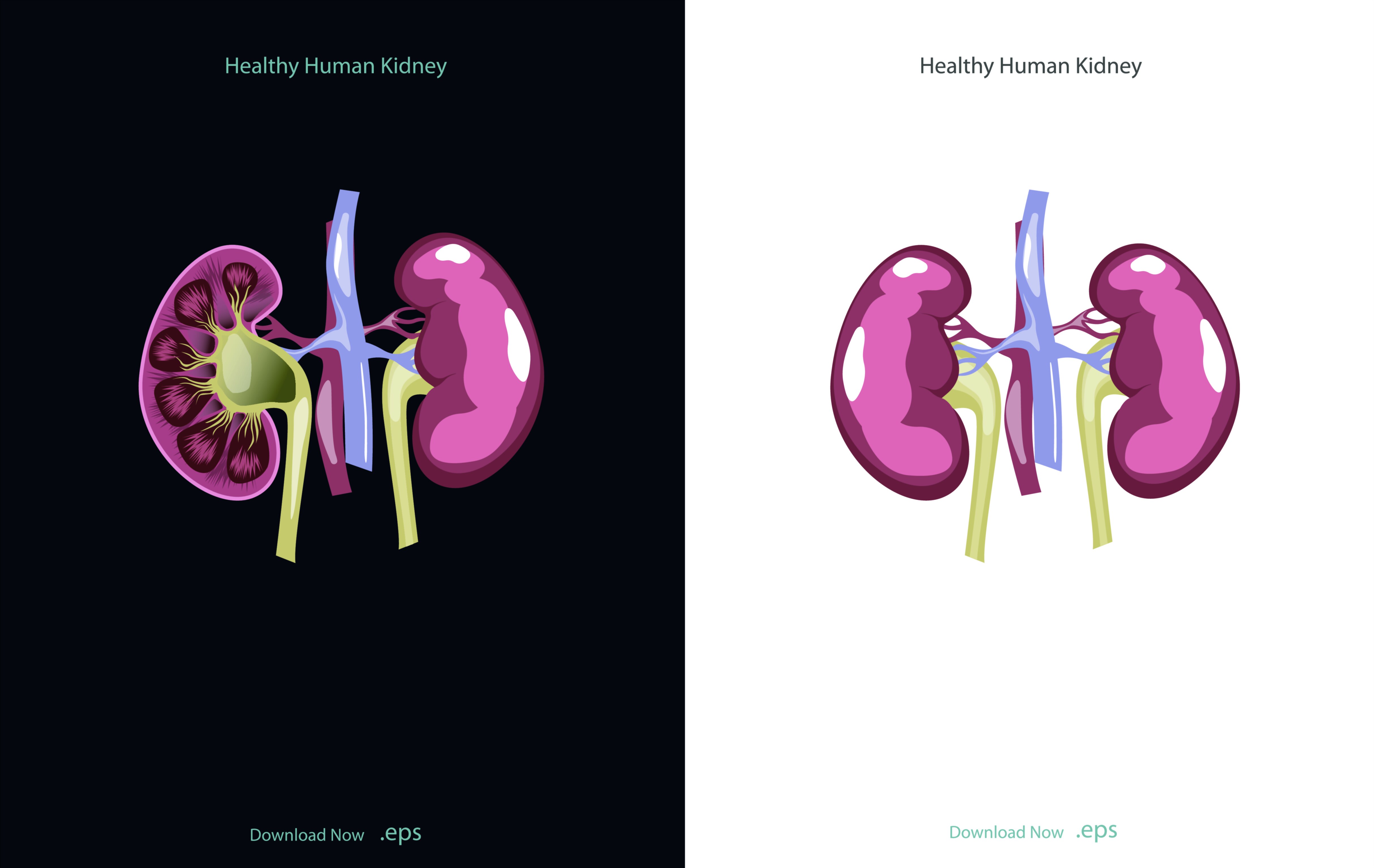
The human kidneys represent one of the most sophisticated biological filtration and regulation systems in the body, combining structural precision with biochemical control to maintain internal balance. Each kidney has a characteristic bean-like shape and is located deep in the posterior abdominal cavity on either side of the spine, protected by muscle, fat, and the lower ribs. Although they are relatively small, they receive nearly a quarter of the blood pumped by the heart because the body relies on constant filtration to remove metabolic waste, regulate water and mineral levels, balance pH, and maintain blood pressure. A vector illustration of kidney structure typically highlights the outer cortex, the inner medulla organized into renal pyramids, the renal pelvis, and the ureter that carries urine toward the bladder, along with blood vessels entering through the renal artery and exiting through the renal vein. These visual elements form a complete map of how the kidney’s physical organization supports its function.
Inside the kidney, the outer renal cortex acts as the entry point for blood filtration. Here lie the glomeruli and the initial segments of microscopic filtration units called nephrons. Each nephron begins with a glomerulus—a tangle of fine capillaries where blood pressure forces plasma out of the bloodstream and into Bowman’s capsule. This marks the beginning of the urine-forming process not through waste expulsion but by separating the liquid portion of blood from its cells and large proteins. The filtered fluid then travels along a specialized tubular pathway through the proximal tubule, the loop of Henle, the distal tubule, and finally the collecting duct. Along this journey, useful substances—like water, glucose, sodium, and amino acids—are reabsorbed back into the bloodstream, while metabolic waste products, toxins, and excess ions remain in the filtrate to become urine. A kidney illustration showing these tubular structures helps learners understand that urine is not stored blood waste but the result of precise sorting, filtering, reabsorbing, and secretory processes.
The renal medulla contains the loop of Henle and collecting ducts arranged in triangular renal pyramids. Their design creates osmotic gradients that allow the kidney to produce urine of varying concentration depending on the body’s hydration state. When water must be conserved, the nephrons reabsorb more water so the body produces small amounts of highly concentrated urine. When excess water must be removed, reabsorption decreases, producing large volumes of dilute urine. This fluid-balancing system is under hormonal control: ADH (antidiuretic hormone) increases water reabsorption, while aldosterone influences sodium and potassium regulation. These endocrine controls highlight that the kidney is not merely a filter—it is a dynamic regulator influencing blood pressure, hydration, and electrolyte equilibrium every moment.
Another essential function of the kidneys is maintaining acid–base balance. Cells throughout the body depend on a stable pH to function properly, and the kidneys actively regulate it by secreting or reabsorbing hydrogen and bicarbonate ions in the nephron. This system works hand-in-hand with the lungs, which control carbon dioxide levels; together they form a dual mechanism that keeps the blood within the narrow pH range necessary for life. Without the kidney’s long-term chemical adjustments, even slight pH disruptions would destabilize metabolic processes.
The kidneys also play a decisive role in blood pressure regulation via the renin–angiotensin–aldosterone system (RAAS). When blood pressure drops or sodium levels decrease, the kidneys release renin, initiating a chain reaction that causes the constriction of blood vessels and increases sodium and water retention to restore circulation. This makes the kidneys central to cardiovascular health. Because of this connection, chronic kidney issues can contribute to hypertension, and uncontrolled high blood pressure can damage kidney tissues in return—creating a biological feedback loop.
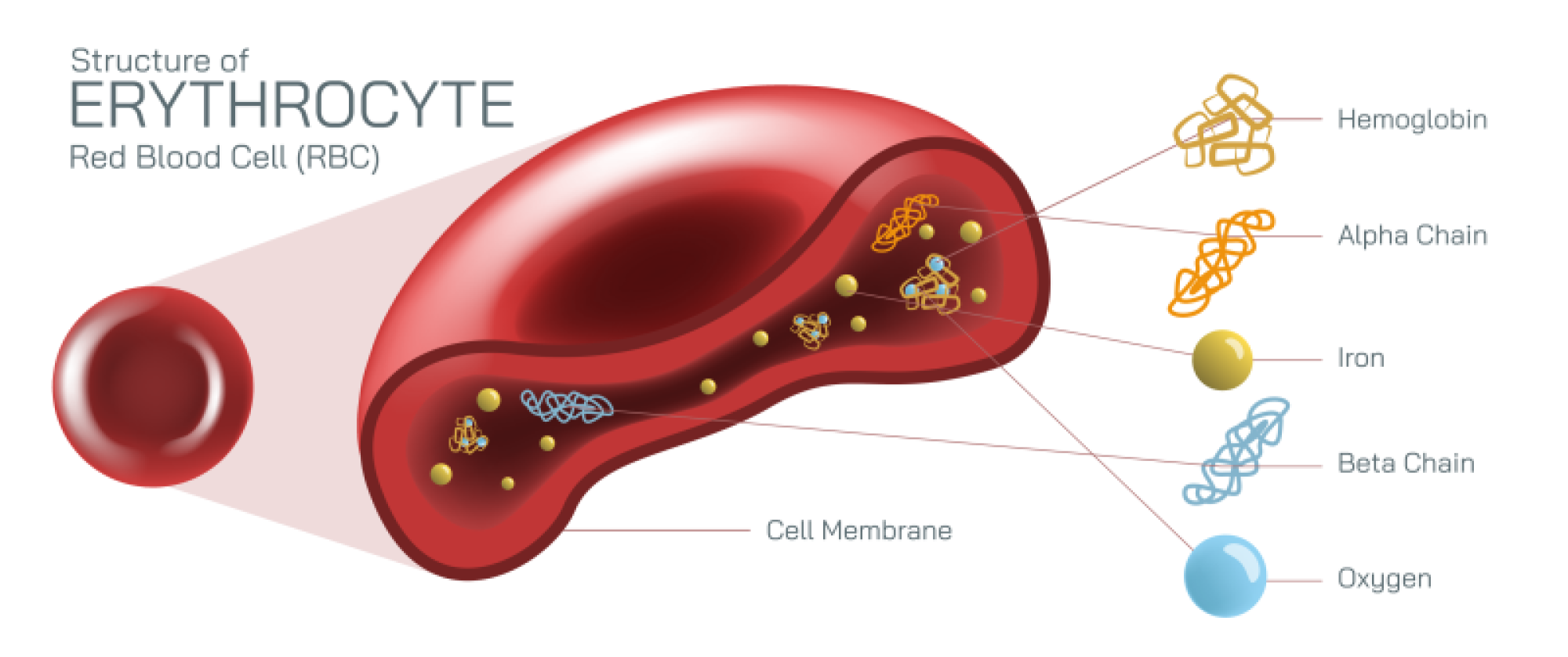
Beyond filtration and regulation, the kidneys are significant endocrine organs. They release erythropoietin, which stimulates the bone marrow to produce red blood cells, connecting renal health to oxygen delivery throughout the body. The kidneys also activate vitamin D, enabling the intestines to absorb calcium for bone strength. When kidney function declines, anemia and bone weakness often follow—not because the blood or bones fail independently, but because the kidneys no longer send the biochemical signals that sustain them. A vector illustration that includes these hormonal pathways helps learners understand that the kidneys influence the entire body rather than remaining isolated to the urinary system.
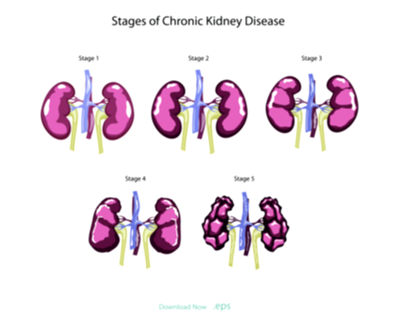
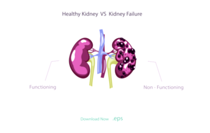
After filtration and regulation, the produced urine drains from the collecting ducts into the renal pelvis and flows through the ureters to the bladder, where it is stored until urination. This removal of waste prevents the buildup of urea, creatinine, and toxins that would otherwise damage tissues and interfere with essential cellular functions. When the kidneys fail, these wastes accumulate, leading to swelling, nausea, fatigue, confusion, and heart strain—illustrating how dependent survival is on continuous renal filtration. Dialysis compensates for failed kidneys by performing artificial filtration, but it can only replace part of the kidneys’ broad regulatory capacity, showing how extraordinary the natural organs are.
A vector illustration of kidney structure brings all of these concepts together visually, showing how the cortex, medulla, pyramids, pelvis, and blood vessels operate as a coordinated filtration and regulation system. It allows students to map the path of blood, the movement of filtrate, the return of vital substances, and the elimination of waste—all in a single design. The kidneys demonstrate that homeostasis, the balance that keeps the body alive, does not emerge from stability but from constant adjustment. Through filtration, reabsorption, hormonal control, pH balance, pressure regulation, and endocrine signaling, the kidneys maintain the internal environment that sustains every organ and every cell.
Ultimately, the human kidney represents a biological masterpiece of precision and adaptability. It works continuously, silently, and without conscious awareness, yet it protects life every moment by purifying blood, conserving resources, eliminating harmful substances, and orchestrating chemical balance. Understanding kidney structure and function through a vector illustration turns anatomy into insight: the shape of each region exists for a purpose, each tubule supports a vital step, and the entire organ serves as the guardian of the body’s internal equilibrium.