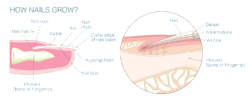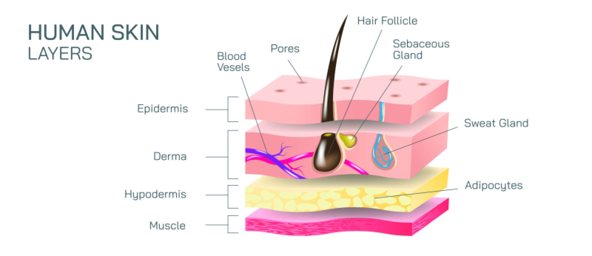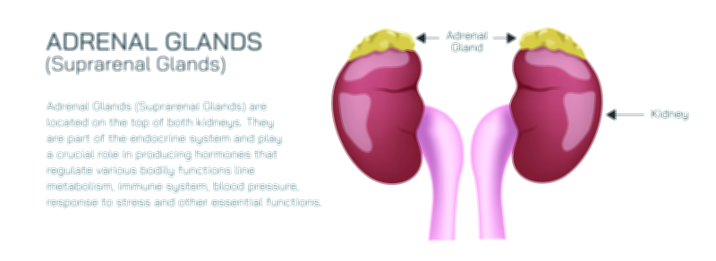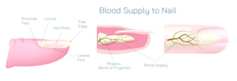
Water in the Human Body Vector Illustration Showing Distribution and Vital Roles in Organs and Cells
Water is the most abundant and vital molecule in the human body, constituting approximately 60% of total body weight in adults and playing a critical role in maintaining physiological balance, cellular function, and systemic homeostasis. A vector illustration depicting water in the human body typically emphasizes the distribution of water across compartments, including intracellular, extracellular, interstitial, and plasma components, as well as its functional roles in organs and cells. By combining anatomical visualization with physiological context, such illustrations provide a clear and educational overview of how water sustains life, facilitates biochemical processes, and supports structural and functional integrity in humans.
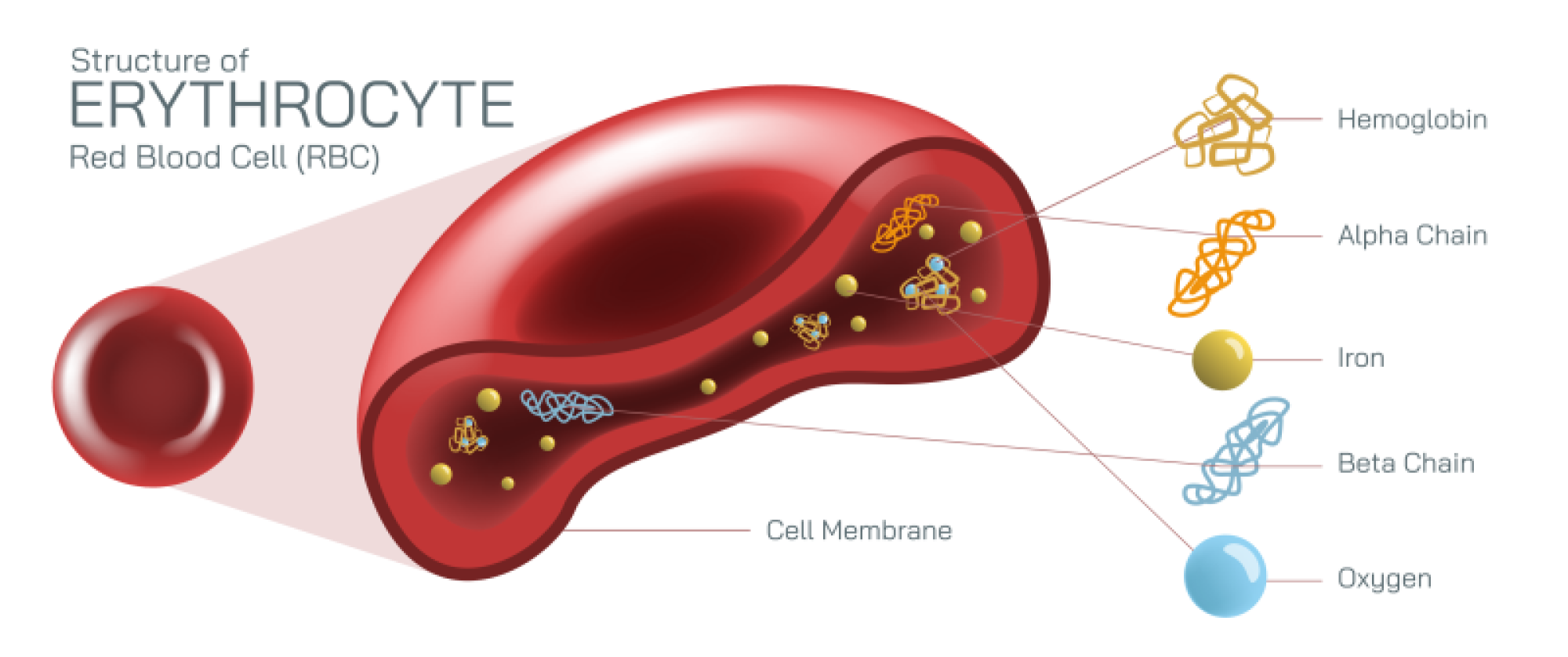
At the core of the illustration is the overall water distribution in the human body. Water is present in intracellular compartments, constituting roughly two-thirds of total body water, and is essential for maintaining cell turgor, biochemical reactions, and nutrient transport. Vector diagrams often highlight the intracellular space within representative cells, using shading or color coding to indicate water content and demonstrating that water forms the medium in which metabolic reactions occur. Extracellular water, making up about one-third of total body water, is subdivided into interstitial fluid surrounding cells, and plasma, the liquid component of blood. Vector illustrations often depict fluid compartments with arrows showing movement between cells, interstitial spaces, and the circulatory system, emphasizing the dynamic equilibrium of water distribution.
The plasma compartment is particularly significant for maintaining blood volume, pressure, and nutrient transport. In vector diagrams, plasma is often illustrated as a component of the circulatory system, highlighting veins, arteries, and capillaries. Arrows may indicate the flow of water between plasma and interstitial fluid through capillary walls via osmosis and filtration, demonstrating the principles of fluid balance and homeostasis. Similarly, the interstitial fluid surrounding cells is depicted as a bridge between plasma and intracellular compartments, facilitating exchange of nutrients, waste products, and signaling molecules. By visualizing these fluid dynamics, the illustration conveys how water maintains tissue hydration, nutrient supply, and cellular function.
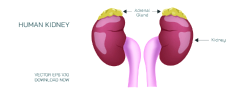
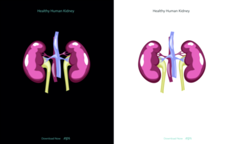
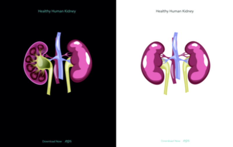
Vector illustrations often emphasize the functional roles of water in various organs and systems. In the kidneys, water is crucial for urine formation, osmoregulation, and electrolyte balance. Arrows may depict filtration in nephrons, showing water movement across membranes and its role in excreting metabolic waste while conserving essential ions. In the digestive system, water aids in digestion, absorption, and transport of nutrients, with arrows showing its involvement in chyme formation and nutrient solubilization. In the circulatory system, water maintains blood viscosity, enables transport of oxygen and nutrients, and facilitates thermoregulation. By labeling organ-specific roles, the illustration highlights how water is integral to both structural and functional processes across the body.
Cellular functions of water are also highlighted in vector illustrations. Water acts as a solvent for biochemical reactions, participates in hydrolysis and condensation reactions, maintains cell shape through osmotic pressure, and facilitates intracellular transport of ions and molecules. Shading or highlighting within a cell may indicate cytoplasmic water, vesicle transport, or organelle hydration, illustrating the concept that water is essential for proper cellular physiology. The illustration may also depict water in organelles like the nucleus and mitochondria, emphasizing its role in energy production, genetic regulation, and metabolic processes.
Vector diagrams often include fluid balance and homeostasis mechanisms, showing how the body regulates water intake and output. Arrows may indicate ingestion from food and beverages, absorption in the gastrointestinal tract, distribution to cells and interstitial spaces, and loss through urine, sweat, respiration, and feces. Labels may highlight regulatory organs and hormones such as the kidneys, hypothalamus, antidiuretic hormone (ADH), and aldosterone, demonstrating how water balance is maintained in response to environmental and physiological changes. This dynamic representation reinforces the concept of homeostasis, showing how water is continuously monitored and adjusted to sustain life.
Illustrations may also depict water’s role in temperature regulation, showing sweat glands and evaporative cooling mechanisms, or its involvement in lubrication and cushioning, highlighting joints, cerebrospinal fluid, and amniotic fluid. By including multiple systems, the diagram demonstrates that water is not confined to a single function but is an omnipresent component facilitating structural support, biochemical reactions, nutrient transport, and protective functions throughout the body.
Color coding, arrows, and labeled compartments in vector illustrations provide clarity by distinguishing intracellular vs. extracellular fluid, highlighting organ-specific functions, and visualizing the dynamic movement of water throughout the body. Magnified insets of cells, kidneys, or joints can provide detail on micro-level processes while retaining an overall systemic perspective. Side-by-side comparisons of fluid distribution in different body compartments may further illustrate proportional contributions to total body water.
By integrating anatomical, cellular, and functional perspectives, a vector illustration of water in the human body provides a comprehensive understanding of its essential roles. It conveys how water supports cellular metabolism, organ function, thermoregulation, nutrient transport, and systemic homeostasis, linking microscopic and macroscopic processes. The diagram allows viewers to appreciate both the quantitative distribution of water and its qualitative significance in sustaining life.
Ultimately, a vector illustration of water in the human body demonstrates the centrality of water in human physiology. By visually representing its distribution across intracellular and extracellular compartments, highlighting organ-specific functions, and depicting its role in cellular and systemic processes, the diagram provides an educational tool that integrates structure, function, and dynamics. Through labeled compartments, arrows, and functional annotations, it transforms an abstract concept into a tangible framework, helping learners and healthcare professionals understand the vital importance of water in maintaining life, health, and physiological balance.