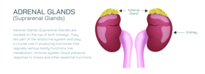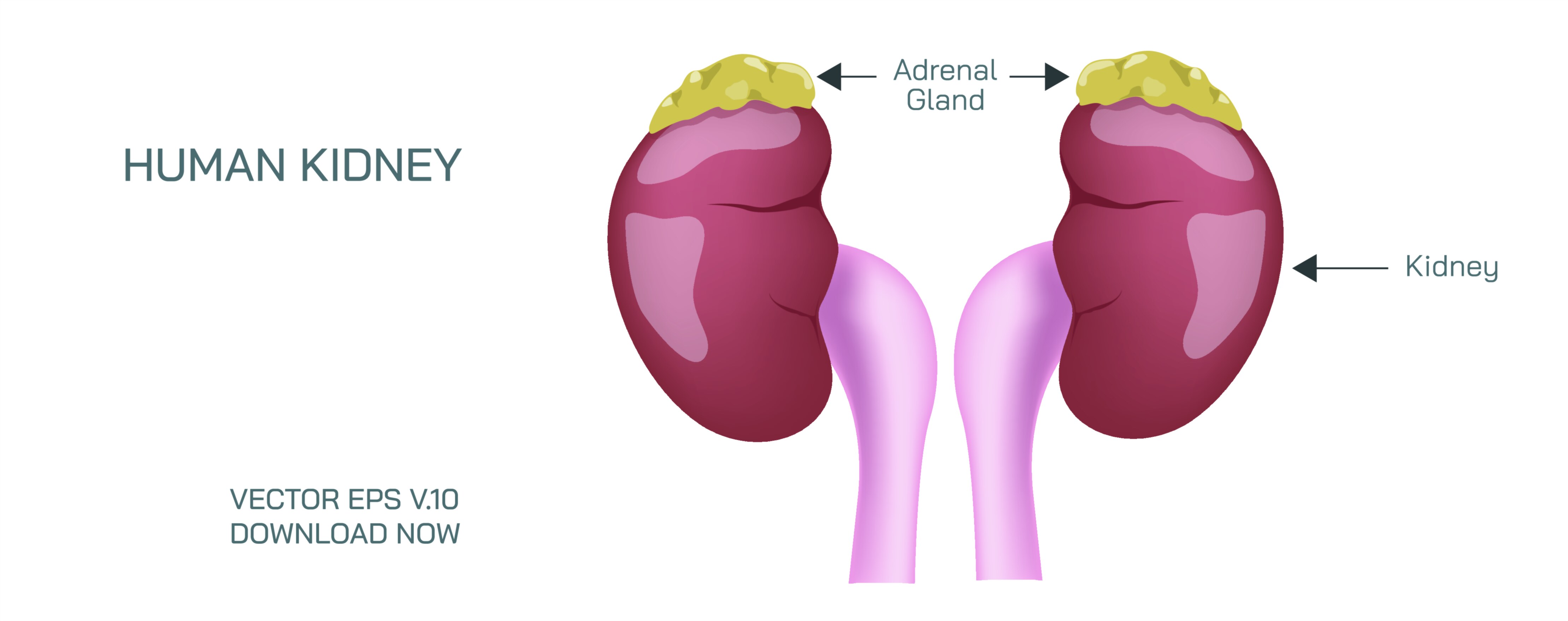
Human Kidney and Adrenal Gland Vector Illustration Showing Structure and Function in Urinary and Endocrine System
The human kidney and adrenal gland are paired organs situated in the retroperitoneal space of the upper abdomen, each performing distinct yet interconnected functions essential for maintaining homeostasis. The kidney is primarily responsible for filtration of blood, waste excretion, electrolyte balance, and fluid regulation, while the adrenal gland, located atop the kidney, produces hormones critical for metabolism, stress response, and electrolyte homeostasis. A vector illustration depicting the kidney and adrenal gland typically highlights their anatomical structures, internal components, and functional roles, providing a comprehensive educational tool for understanding the urinary and endocrine systems. By integrating visual anatomy with functional context, the diagram elucidates how these organs interact to support systemic physiology.
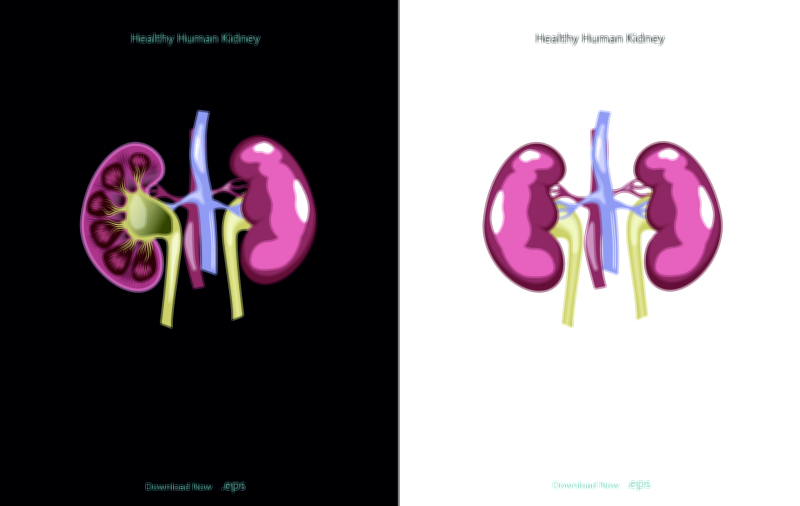
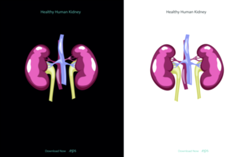
The kidney anatomy is a central focus of the illustration. The kidney is typically shown in a bean-shaped form, with external and internal structures labeled. The outer layer, the renal cortex, contains the glomeruli and proximal and distal convoluted tubules, while the inner layer, the renal medulla, houses the renal pyramids, collecting ducts, and portions of the nephron responsible for urine concentration. Vector diagrams often depict a cross-sectional view, showing the cortex, medulla, renal pelvis, and calyces, with arrows indicating the flow of filtrate from nephrons to the ureter. The renal pelvis, a funnel-shaped structure at the hilum, collects urine and channels it into the ureter, which is also labeled in the illustration. Color coding or shading may be used to differentiate functional regions, emphasizing the structural organization that supports filtration, reabsorption, and excretion.
A critical component in kidney illustrations is the nephron, the functional unit responsible for filtering blood and forming urine. Vector diagrams often include a magnified nephron inset, labeling the glomerulus, Bowman's capsule, proximal tubule, loop of Henle, distal tubule, and collecting duct. Arrows may indicate the directional flow of filtrate and the selective reabsorption and secretion of water, ions, and waste products. Although chemical equations are not displayed, the diagram visually conveys the movement of fluids and solutes, demonstrating how the nephron maintains fluid and electrolyte balance, removes metabolic wastes, and contributes to blood pressure regulation through the renin-angiotensin-aldosterone system (RAAS).
The adrenal glands, perched atop each kidney, are represented as small, triangular organs. Each adrenal gland is divided into the cortex and medulla, with vector illustrations labeling the distinct layers of the cortex—zona glomerulosa, zona fasciculata, and zona reticularis—and the central medulla. The cortex is responsible for producing steroid hormones, including mineralocorticoids (aldosterone) for sodium and water balance, glucocorticoids (cortisol) for metabolic regulation, and androgens. The medulla secretes catecholamines, including epinephrine and norepinephrine, essential for the body’s stress response and sympathetic nervous system activation. Arrows and labels in vector illustrations often indicate hormone secretion pathways from the adrenal gland into the bloodstream, demonstrating the endocrine function in parallel with the kidney’s excretory role.
Vector illustrations may also highlight the renal vasculature, showing how blood enters the kidney via the renal artery, branches into afferent arterioles supplying the glomeruli, and exits through the renal vein. Arrows may indicate blood flow through the nephron, emphasizing filtration at the glomerulus and reabsorption along the tubules. The diagram may also show the close association of adrenal blood supply, highlighting how adrenal arteries branch from the renal artery or directly from the aorta. This vascular depiction illustrates the integration of urinary and endocrine function, as blood supply delivers substrates for filtration, oxygenation, and hormone transport.
Additional elements in vector diagrams may include urinary system connections, such as ureters, bladder, and urethra, situating the kidney within the broader excretory pathway. Similarly, the adrenal gland’s relation to the sympathetic nervous system can be illustrated with arrows indicating neural inputs that stimulate catecholamine release during stress responses. Color coding may differentiate kidney structures, adrenal layers, vasculature, and urine flow, enhancing visual clarity and learning.
Functional annotations in the illustration may highlight key physiological processes, such as glomerular filtration, tubular reabsorption, urine concentration, hormone secretion, and regulation of blood pressure and fluid balance. Arrows may indicate water and solute movement in the nephron, while labels identify hormone types and target organs for adrenal secretions. These features integrate anatomical detail with physiological function, emphasizing how the kidney and adrenal gland work in tandem to maintain homeostasis.
By combining kidney and nephron structure, adrenal gland layers, vascular supply, and functional flow pathways, a vector illustration provides a detailed and holistic view of urinary and endocrine physiology. The diagram demonstrates the interdependence of filtration, hormone production, fluid balance, and systemic regulation. Magnified insets, cross-sectional views, and labeled arrows enhance comprehension, showing the dynamic processes occurring within and between these organs.
Ultimately, a vector illustration of the human kidney and adrenal gland conveys the structural complexity and functional integration of these vital organs. Through labeled anatomy, visual flow of urine and hormones, and clear depiction of tissue layers, the diagram transforms complex biological systems into an accessible, educational, and visually intuitive framework, enabling learners and healthcare professionals to understand the critical roles of the kidney and adrenal gland in maintaining human health.