Adrenal Gland Template Showing Structure, Hormone Zones, Cortex, and Medulla for Medical Study
The adrenal glands are small, triangular endocrine organs positioned above the kidneys, yet their internal structure and hormonal output make them some of the most influential regulators of stress response, metabolism, blood pressure, immune function, and overall physiological balance. A medical study template that clearly illustrates the structure of the adrenal glands—with emphasis on their outer cortex, inner medulla, and the three distinct hormone-producing zones—provides an invaluable foundation for learning endocrine physiology. By organizing the anatomy visually and conceptually, students can understand not only where different hormones come from but why each region is specialized for its own regulatory role. Although the glands are only a few centimeters in size, their layered structural design enables them to mobilize rapid emergency response while simultaneously sustaining long-term metabolic and homeostatic regulation.
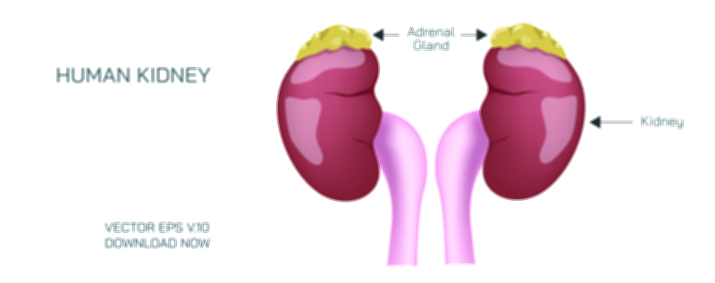
The template begins with the external appearance of each adrenal gland, which resembles a flattened pyramid on the right kidney and a crescent shape on the left kidney. A thin capsule wraps the organ, but immediately beneath this surface lies the adrenal cortex, a golden-brown tissue that makes up roughly 80–90% of the gland’s mass. The cortex is subdivided into three well-defined hormonal layers organized like concentric shells, each responsible for producing a distinct category of steroid hormones. The organization of these zones is not simply anatomical; it reflects biochemical specialization rooted in enzyme expression and signaling pathways. The medical template illustrates these with clear boundaries so that the function of the cortex is taught as a layered progression rather than a single mass of endocrine tissue.
The outermost layer, the zona glomerulosa, appears as a thin band beneath the capsule. It is the exclusive site of synthesis for mineralocorticoids, primarily aldosterone. These hormones regulate sodium and potassium levels in the bloodstream, influence water retention, and directly affect blood pressure. The zona glomerulosa responds mainly to signals from the renin-angiotensin-aldosterone system, making it essential to cardiovascular and renal physiology. In a classroom template, this outer band is typically highlighted first, emphasizing its unique function and its conversation with the kidneys and circulatory system.
Beneath it lies the zona fasciculata, the thickest region of the adrenal cortex and one of the most visually distinctive in histological diagrams because of its vertically arranged columns of cells. This middle layer produces glucocorticoids, the most clinically significant being cortisol, which plays an enormous role in glucose metabolism, immune modulation, inflammation control, protein and fat mobilization, and long-term stress adaptation. This zone interacts closely with the hypothalamic-pituitary-adrenal (HPA) axis, responding to ACTH released from the anterior pituitary. When the adrenal gland template is used in medical study, the zona fasciculata is often placed at the conceptual center of adrenal physiology because disturbances in cortisol secretion—whether deficiency or excess—have wide-ranging effects across nearly every organ system.
The innermost cortical layer, the zona reticularis, sits between the zona fasciculata and the medulla and synthesizes adrenal androgens, including dehydroepiandrosterone (DHEA) and androstenedione. Although these hormones are weaker than the primary sex steroids produced in the gonads, they contribute to sexual development, puberty, libido, and overall energy and mood regulation. In women, adrenal androgens represent an especially important source of testosterone precursors. A detailed template presents this inner layer with particular clarity because students often overlook its significance compared to aldosterone and cortisol, even though it provides essential hormonal support during adolescence and aging.
At the absolute center of the adrenal gland sits the adrenal medulla, structurally and functionally distinct from the cortex. Derived from neural crest tissue rather than mesoderm, the medulla operates as a modified sympathetic ganglion, releasing catecholamines—primarily epinephrine (adrenaline) and norepinephrine (noradrenaline)—directly into the bloodstream. These hormones fuel the body’s fight-or-flight response, dramatically increasing heart rate, respiratory function, mental alertness, glucose availability, and muscular readiness in response to sudden threat or intense stress. A visual template that separates the medulla from the cortex reinforces that adrenal physiology works on two scales simultaneously: the cortex supports sustained homeostasis and adaptation, while the medulla enables rapid emergency activation.
Because the adrenal glands rely on multiple signaling systems, an effective medical illustration often includes directional arrows and brief labeling to show the regulatory pathways. The HPA axis controls cortisol release through hypothalamic CRH and pituitary ACTH, while the renin-angiotensin-aldosterone system regulates mineralocorticoid activity. Sympathetic neurons activate catecholamine release from the medulla nearly instantaneously. When these signaling networks are represented visually together with the cortex and medulla, the student begins to appreciate the adrenal glands not as simple hormone reservoirs but as highly responsive communication hubs linking the brain, kidneys, immune system, metabolism, and cardiovascular system.
The physiological implications of this organization become even more striking when learning about disorders. Because each layer of the adrenal gland produces different hormones, dysfunction presents in distinct patterns. Insufficient cortisol and aldosterone, as in Addison’s disease, manifests with chronic fatigue, dehydration, low blood pressure, weight loss, and electrolyte imbalance. Excess cortisol, as in Cushing’s syndrome, leads to central weight gain, muscle wasting, fragile skin, elevated blood sugar, and immune suppression. Mineralocorticoid-producing tumors cause severe hypertension, while medullary tumors (pheochromocytomas) can produce episodic surges of adrenaline that mimic panic attacks, tachycardia, and dangerously high blood pressure. When a template labels dysfunction by zone, the relationship between hormone and symptom becomes clear and clinically memorable.
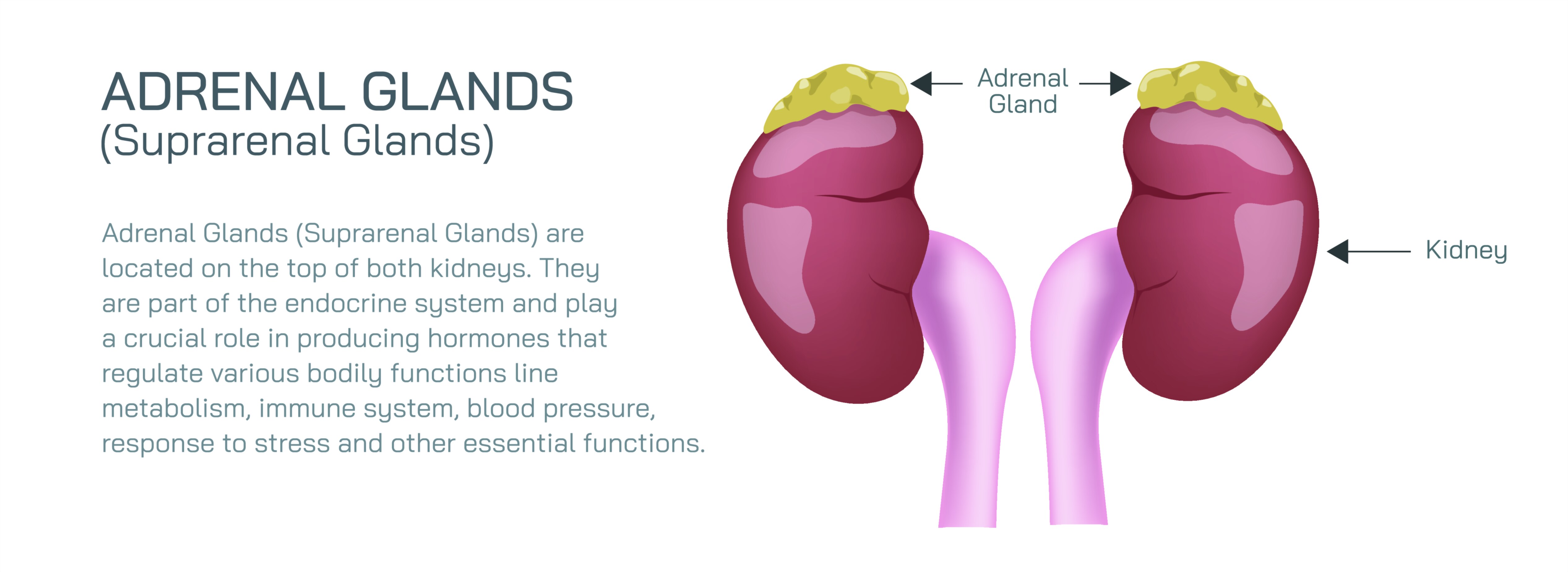
A medical study diagram of the adrenal gland that shows structural boundaries—capsule, three cortical zones, and central medulla—therefore does more than list anatomy. It teaches an organizational logic: structure determines hormone type, and hormone type determines physiological effect. By anchoring the adrenal glands visually above the kidneys, labeling each production zone distinctly, separating cortex from medulla, and connecting each region to its endocrine roles, the template becomes an essential educational map of one of the most sophisticated hormone-producing organs in the body. It clarifies why adrenal health is a pillar of biological resilience and why disruption in any of its zones affects energy, immunity, stress tolerance, metabolism, hydration, cardiovascular health, and overall homeostasis.