Human Kidney Anatomy and Function — Structure, Filtration, and the Body’s Internal Balance System
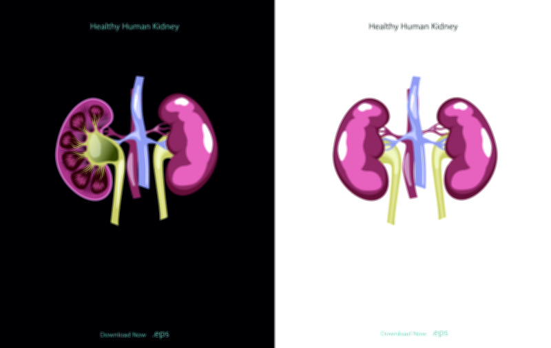
The human kidneys are among the most vital organs for sustaining life because they continuously filter the blood, remove metabolic waste, regulate water and electrolyte balance, maintain pH, and control blood pressure through an intricate biological system that operates every second without conscious awareness. Each kidney is shaped like a bean and positioned deep in the abdominal cavity on either side of the spine, protected by muscles and ribs. Although small and lightweight compared to many internal organs, the kidneys receive an astonishingly large portion of the body’s blood flow—nearly 20–25% of total cardiac output—because their function depends on processing vast amounts of blood to maintain a stable internal environment. A vector illustration of kidney anatomy typically highlights the cortex, medulla, renal pyramids, renal pelvis, and the ureter, along with branching blood vessels entering through the renal artery and exiting through the renal vein. Such a diagram provides a structural window into one of the body’s most advanced biological filtration systems.
The renal cortex, the outermost region, contains dense networks of capillaries and the beginning sections of the nephron—the microscopic functional units responsible for filtration. Moving inward toward the renal medulla, the viewer encounters the renal pyramids, where tubular structures extend and concentrate urine through fluid and solute exchanges, all controlled by gradient differences created by sodium and other electrolytes. At the deepest part lies the renal pelvis, a collection basin where newly formed urine gathers before passing into the ureter. A vector illustration that labels these parts helps learners visualize how structure supports function: the outer cortex initiates filtration, the inner medulla concentrates urine, and the pelvis channels waste downward for elimination.
The nephron is the true engine of kidney function. Each kidney contains roughly one million nephrons, and each nephron begins with a glomerulus, a cluster of capillaries through which blood plasma is filtered into the Bowman’s capsule. The filtered fluid then travels through the proximal tubule, loop of Henle, distal tubule, and collecting duct, undergoing selective reabsorption and secretion along the way. This means the kidney does not simply push wastes out—it separates useful substances from harmful ones, reclaiming water, glucose, amino acids, and electrolytes to return to the bloodstream while allowing urea, creatinine, toxins, drugs, and excess ions to leave the body as urine. A kidney illustration showing this tubular pathway clarifies that urine is not pre-made waste but the result of continuous biochemical sorting controlled by hormones and cellular transport.
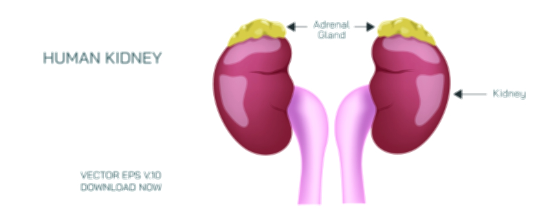
The kidneys play a commanding role in fluid balance. When the body holds too much water, the kidneys respond by excreting dilute urine; when water intake is low or sweating is excessive, the kidneys conserve water by producing highly concentrated urine. This adaptability is guided by hormones such as ADH (antidiuretic hormone), which increases water reabsorption, and aldosterone, which regulates sodium and potassium balance. These hormonal interactions help the body maintain stable blood pressure and prevent dangerous fluctuations in electrolytes. A vector diagram showing hormone influence across nephron segments can help students understand how the endocrine system integrates with renal function.
Another essential responsibility of the kidneys is the regulation of acid–base balance, maintaining the blood’s pH within the narrow range required for cellular function. Through the secretion or reabsorption of hydrogen and bicarbonate ions, the kidneys act as a long-term stabilizing system, correcting imbalances that the lungs alone cannot manage. Without this ongoing fine-tuning, enzymes would cease to function properly, metabolic reactions would fail, and the body’s internal chemistry would fall out of equilibrium.
The kidneys are also key organs in blood pressure regulation through the renin–angiotensin–aldosterone system (RAAS). When blood pressure drops, the kidneys release renin, setting off a cascade of reactions that constrict blood vessels and regulate salt and water retention. This mechanism ensures that tissues receive adequate blood flow even during dehydration or injury. An anatomical illustration that connects the renal artery to systemic circulation helps depict the kidneys not merely as filters but as regulators of cardiovascular stability.
In addition to filtration and regulation, the kidneys perform crucial endocrine functions. They produce erythropoietin, a hormone that stimulates bone marrow to create red blood cells, linking renal function to oxygen transport in the body. They also convert vitamin D into its active form, which supports calcium absorption and bone health. When kidney function declines, anemia and weakened bones can develop—not because the digestive or skeletal systems fail, but because the kidneys no longer support them hormonally. A vector illustration that includes endocrine pathways highlights this lesser-known but vital dimension of kidney physiology.
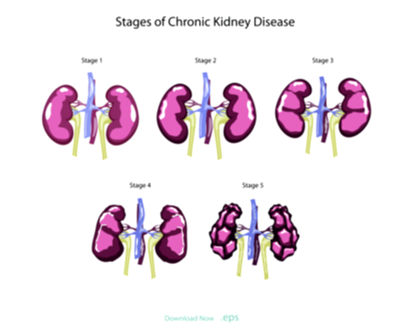
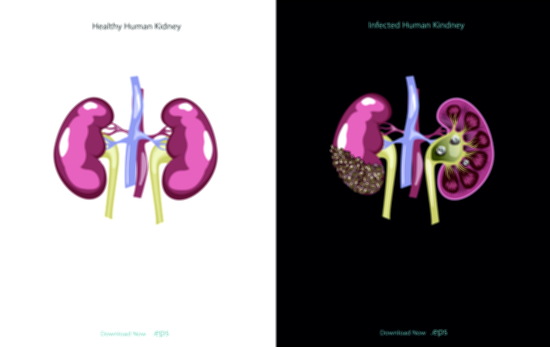
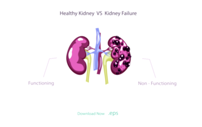
As urine forms, it travels from the collecting ducts into the renal pelvis and then through the ureters to the bladder, where it is stored until urination. This eliminates metabolic waste and prevents the accumulation of toxic substances that would otherwise poison tissues. The kidneys therefore function not only as waste-removal organs but as guardians that maintain internal chemical stability every moment of life. When filtration slows due to disease or injury, the buildup of waste can lead to fatigue, nausea, confusion, swelling, and heart strain. Kidney failure illustrates how dependent human survival is on this filtration process and why dialysis—an artificial method of cleaning the blood—is required when kidneys stop functioning properly.
From an educational perspective, a vector illustration of the kidneys provides a multi-layered learning experience. It reveals how anatomical structures—from cortex to pelvis—support a logical internal flow, how microscopic nephrons perform filtration and reabsorption, how hormonal pathways regulate each step, and how renal health is connected to blood chemistry, skeletal strength, cardiovascular function, and hydration balance. The kidneys symbolize the idea that biological systems maintain life not by keeping things constant, but by continuously adjusting and regulating. Through filtration, regulation, hormone production, and waste elimination, the kidneys sustain the internal environment that every cell in the human body depends on.
Ultimately, human kidney anatomy teaches that life relies on precise balance. The kidneys work silently, without sensation or awareness, yet their continuous activity is as vital as the beating of the heart or the movement of the lungs. Every minute of every day, they monitor the bloodstream, protect against chemical imbalance, and preserve conditions that allow life to flourish. A vector illustration of the kidneys is therefore far more than an anatomical diagram—it is a visual representation of the body’s internal equilibrium, reminding us that longevity and health depend on the unseen but extraordinary work of these remarkable organs.