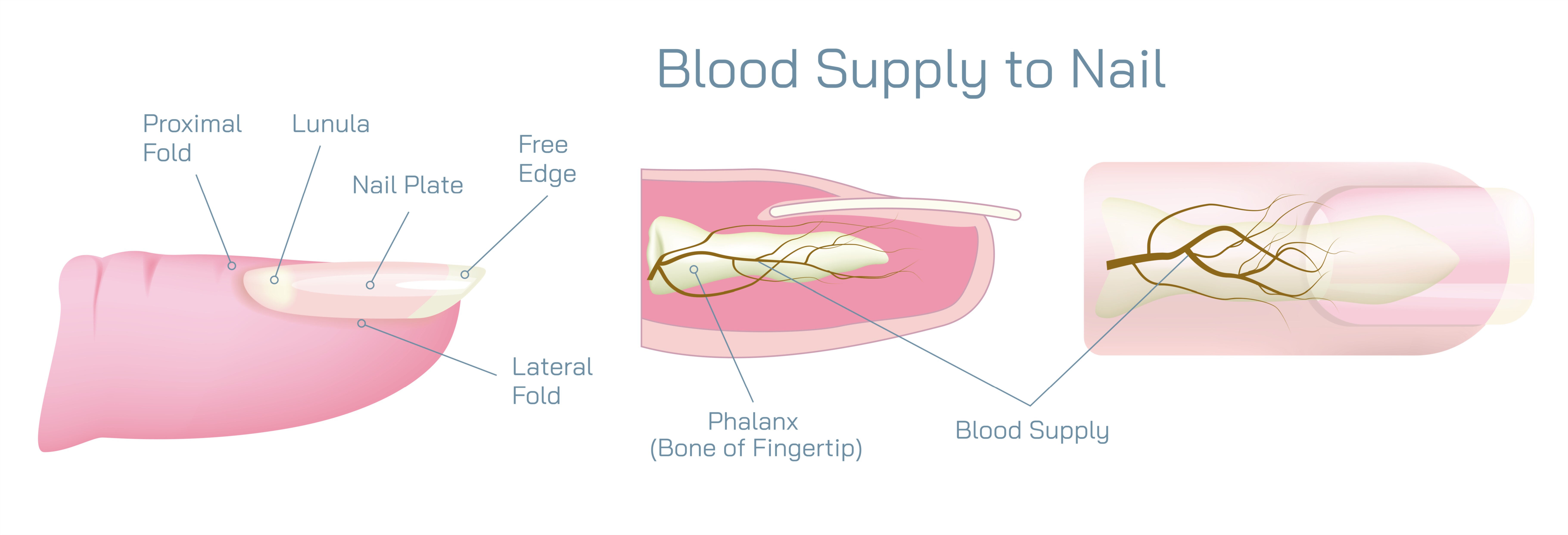
Blood Supply to the Nail Showing Capillaries, Nail Bed Circulation, and Vascular Anatomy in the Human Finger
The nail may appear as a hard, inert surface, but it exists above a highly active and richly supplied vascular system that nourishes the tissues responsible for nail growth and fingertip protection. A vector illustration showing blood supply to the nail typically focuses on how capillaries, nail bed circulation, and vascular anatomy interact beneath the nail plate to sustain the nail unit. This visualization reveals that the nail’s health, color, growth rate, and sensitivity all depend on the underlying blood vessels that feed oxygen and nutrients to surrounding cells.
Directly beneath the nail plate lies the nail bed, a specialized layer of tissue that serves as the primary vascular support system for the nail. This region contains a dense network of tiny blood vessels that run lengthwise under the plate. In an illustration, these capillary loops are often depicted as fine red or blue branches tracking along the nail bed, demonstrating the close contact between the nail surface and circulating blood. Although the nail plate is rigid, it remains thin and semi-transparent, allowing the pink color of the nail bed's blood supply to show through. This is why well-oxygenated blood produces a healthy pink nail appearance, while low oxygen, vascular problems, or poor circulation can make the nail look bluish, pale, or mottled.
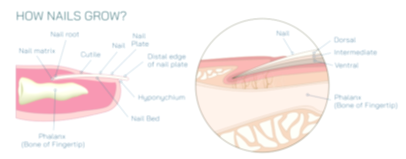
The vascular network of the nail bed delivers continuous nourishment to the tissues surrounding the nail — particularly the matrix, the factory that produces new nail cells. The matrix sits beneath the skin at the base of the nail and depends on robust blood flow to support rapid cell division and keratin formation. Illustrations often show small arteries and venules branching from deeper digital vessels toward the matrix region, emphasizing that nail growth is only possible because the matrix receives a constant supply of oxygen, protein building blocks, vitamins, and minerals through the bloodstream. When blood supply to the matrix is compromised — due to trauma, vascular disease, or systemic illness — nail growth slows, weakens, or becomes irregular, creating ridges or deformities that travel outward over time.
The lunula, the pale crescent visible at the base of some nails, lies above the front portion of the matrix and overlays a transitional vascular area. The lunula appears lighter not because of a lack of blood vessels, but because the semi-keratinized cells in this region are opaque enough to block underlying coloration. In illustrations, the contrast between the pink vascular nail bed and the light lunula helps viewers understand the shift in cell development and optical transparency rather than an absence of circulation.
Beyond the nail bed and matrix, the digital arteries and veins supply the overall fingertip region and feed the smaller vessels supporting the nail. The main blood flow originates from the proper palmar digital arteries, which run alongside the finger on both sides before branching into smaller vessels that extend toward the nail bed and matrix. The venous return follows a similar arrangement, collecting deoxygenated blood and transporting it back toward the hand. A complete anatomical image often shows these vessels running longitudinally along the sides of the digit, highlighting how blood supply wraps around the phalangeal bone to reach the tissue beneath the nail.
Capillary circulation under the nail also plays an important role in thermoregulation and clinical assessment. Because the nail plate is translucent, physicians can observe changes in circulation quickly, such as capillary refill time — the speed at which blood returns to capillaries after pressure is applied. Slow refill may suggest dehydration, shock, or vascular insufficiency. Color changes in the nail bed can also signal anemia, cyanosis, Raynaud’s disease, or systemic illness. These diagnostic uses underscore the importance of the nail’s blood supply not just for nail function, but for evaluating whole-body health.
In addition to nourishment and growth, vascular supply provides sensory support. The fingertip contains an exceptionally high density of nerve endings, and coordinated blood flow helps sustain these sensory neurons. As a result, even though the nail itself has no nerves, the tissue beneath it is extraordinarily sensitive. This heightened sensation allows the nail plate to act as a stiff backing so the fingertip can detect fine textures and pressures with precision.
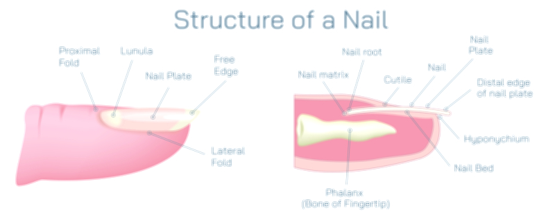
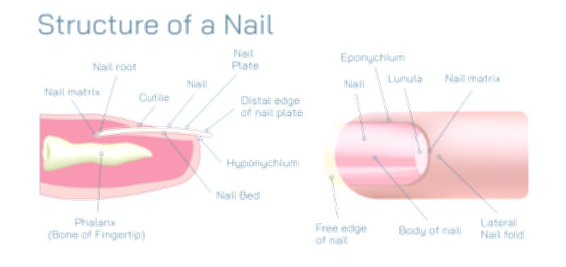
A complete vector illustration of the nail’s blood supply typically includes:
• The nail plate, shown as the semi-transparent hard layer
• The nail bed, illustrated with parallel capillary loops beneath the plate
• The matrix, receiving branching blood vessels supporting nail formation
• The lunula, marking the partially keratinized region overlying transitional vascular structures
• Larger digital arteries and veins, positioned along the sides of the finger and feeding the nail bed circulation
• Arrows or labels to indicate direction of blood flow and key vascular regions
Through the lens of circulation, the nail becomes more than a mechanical shield — it is a biologically supported structure that depends on uninterrupted vascular supply to maintain growth, immunity, coloration, and fingertip sensitivity. The uninterrupted delivery of blood allows the matrix to produce new nail tissue, allows the nail bed to support and anchor the plate firmly, and allows the fingertip to function with remarkable precision and responsiveness.
This intricate vascular anatomy makes the nail a mirror of human health. When circulation is strong, nails grow smoothly and appear vibrant. When blood flow is disrupted, the nail often provides the first visible warning. Illustrating the blood supply beneath the nail — with capillaries, nail bed circulation, and digital vessels working together — exposes the hidden physiological system that keeps every nail alive, growing, and integrated into the broader anatomy of the human finger.