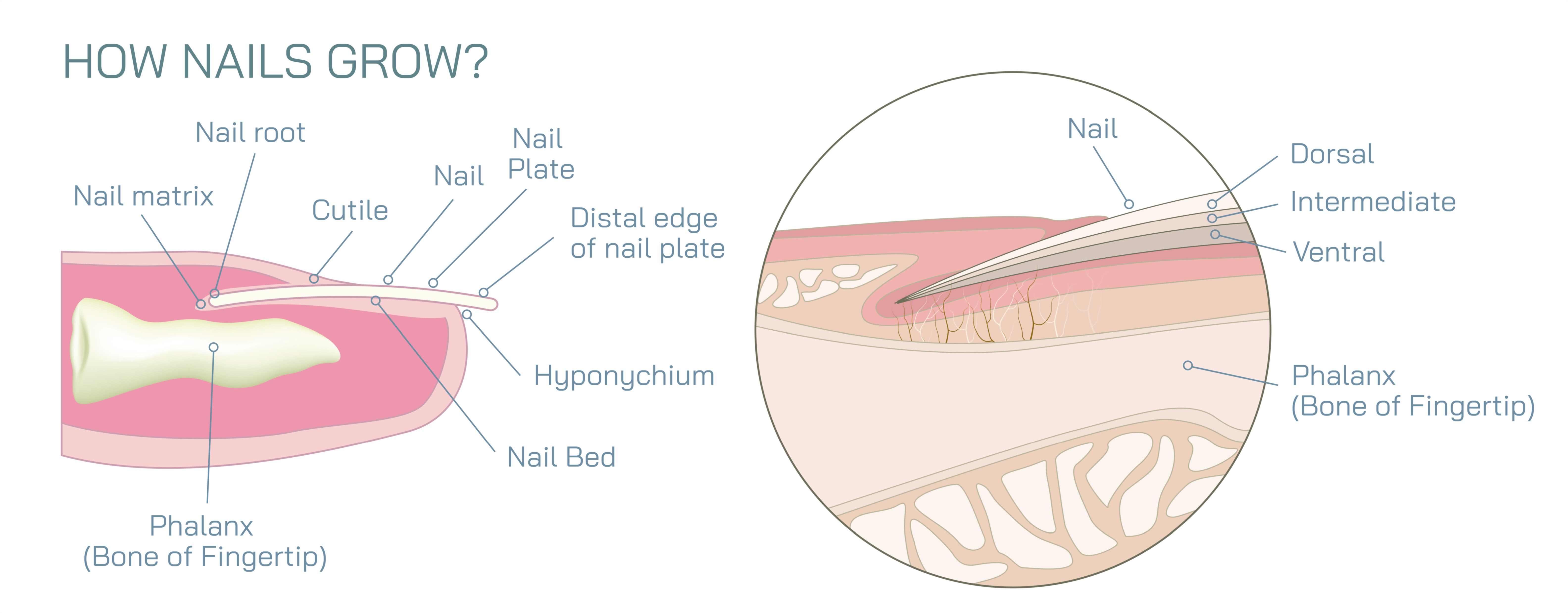
How Nails Grow Showing Nail Matrix, Keratin Formation, and Growth Direction in Human Anatomy
Nails may seem static and unchanging from day to day, but beneath their surface is a highly active biological system that continuously produces new nail material through specialized cellular processes. A vector illustration showing how nails grow usually highlights three key concepts: the nail matrix where growth begins, keratin formation which gives the nail its hardness, and the growth direction that carries the nail plate forward across the fingertip. By visualizing these elements together, the illustration reveals how a living nail is not simply sitting on the finger — it is constantly being regenerated from within.
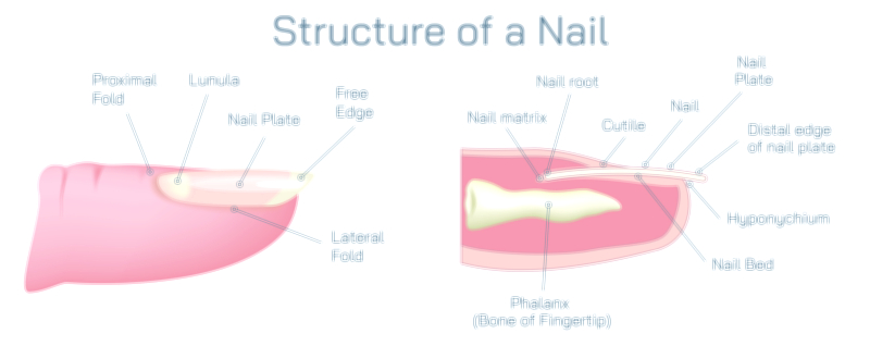
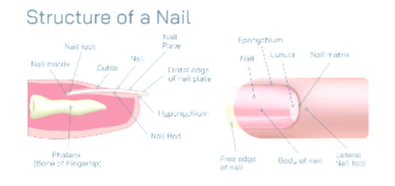
The growth of a nail starts in the nail matrix, the soft tissue hidden beneath the skin at the base of the nail. This region acts as the nail’s production center. Inside the matrix, cells divide rapidly in layers, generating new cells at a pace faster than most other areas of the skin. In a vector diagram, the matrix is located under the proximal nail fold just behind the cuticle, often shown as a crescent-shaped or elongated area responsible for forming the nail plate. Although mostly concealed, part of the matrix is sometimes visible as the pale whitish lunula, the half-moon-shaped area at the base of the nail on some fingers.
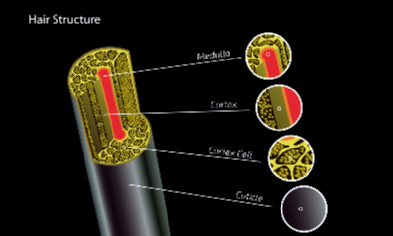
The newly formed cells in the matrix begin as soft, rounded living cells, but as they are pushed forward they undergo keratinization, a transformation that defines nail formation. During keratinization, the cells lose moisture, flatten, and fill with hard keratin, a strong fibrous protein that is also found in hair and the outer layer of skin, but arranged much more densely in nails. Once keratinized, the cells die — not due to damage, but because hardness and inflexibility are necessary for the nail’s protective role. In a vector illustration, this process is often shown as a gradient: soft, living cells in the matrix transitioning to fully hardened and compacted keratin cells as they move outward.
As new keratinized cells accumulate beneath the older ones, the nail plate is gradually pushed forward. This forward motion explains why nails grow outward, not upward or downward. The matrix always produces new nail from behind, and each new cell layer forces the existing nail toward the fingertip. The direction of growth shown in diagrams is usually marked with arrows along the underside of the nail plate, tracing the path from the matrix toward the free edge of the nail. The underside of the plate is anchored to the nail bed, which is why the nail moves as a single continuous sheet rather than lifting off prematurely.
The rate of nail growth varies based on multiple factors — age, nutrition, temperature, circulation, and overall health — but on average, fingernails extend about 3 to 4 millimeters per month, while toenails grow more slowly. Because the nail grows from its base rather than its tip, any injury that affects the matrix affects future growth. Damage to the nail plate itself may cause temporary irregularities, but damage to the matrix can change nail shape, thickness, or growth permanently. Illustrations sometimes emphasize this by showing how trauma to the nail plate does not alter the origin of growth, whereas trauma to the matrix interferes with cell production.
The cuticle also appears in complete diagrams because it protects the top of the matrix. Acting as a natural seal, the cuticle keeps microorganisms from entering the gap where the new nail emerges. Without this protective border, infection or inflammation could reach the matrix and disrupt keratin production. Even though the cuticle does not contribute to nail growth directly, maintaining its integrity preserves the stability of the growth process.
Inside the nail, growth is continuous — there is no shedding phase, unlike hair. The constant creation of keratin from the matrix ensures a steady outward progression. When the nail reaches the tip of the finger, the portion that extends beyond the nail bed becomes the free edge, which eventually wears down or is trimmed but does not influence the pace of growth behind it. The entire visible nail plate therefore represents a timeline of growth: the newest cells are near the cuticle, and the oldest are at the free edge.
A detailed vector illustration of nail growth typically includes:
• The nail matrix as the origin of nail formation
• Newly divided cells becoming progressively keratinized as they move forward
• The nail plate extending outward over the nail bed
• The cuticle protecting the emerging nail at the base
• Arrows indicating the growth direction from matrix to free edge
These components capture how the nail behaves as both a protective tool and a constantly renewing tissue. What looks like a solid, unchanging surface is actually the product of an ongoing biological sequence driven by cell division, keratin production, and forward displacement. By showing the invisible mechanics beneath the visible nail, the illustration provides insight into why nails grow the way they do — steadily, directionally, and dependently on the health of the matrix and its keratin-forming machinery.
At its core, nail growth reflects a remarkably well-orchestrated system: the matrix builds, keratin strengthens, and growth motion advances — all combining to create the durable and functional nail we rely on every day.