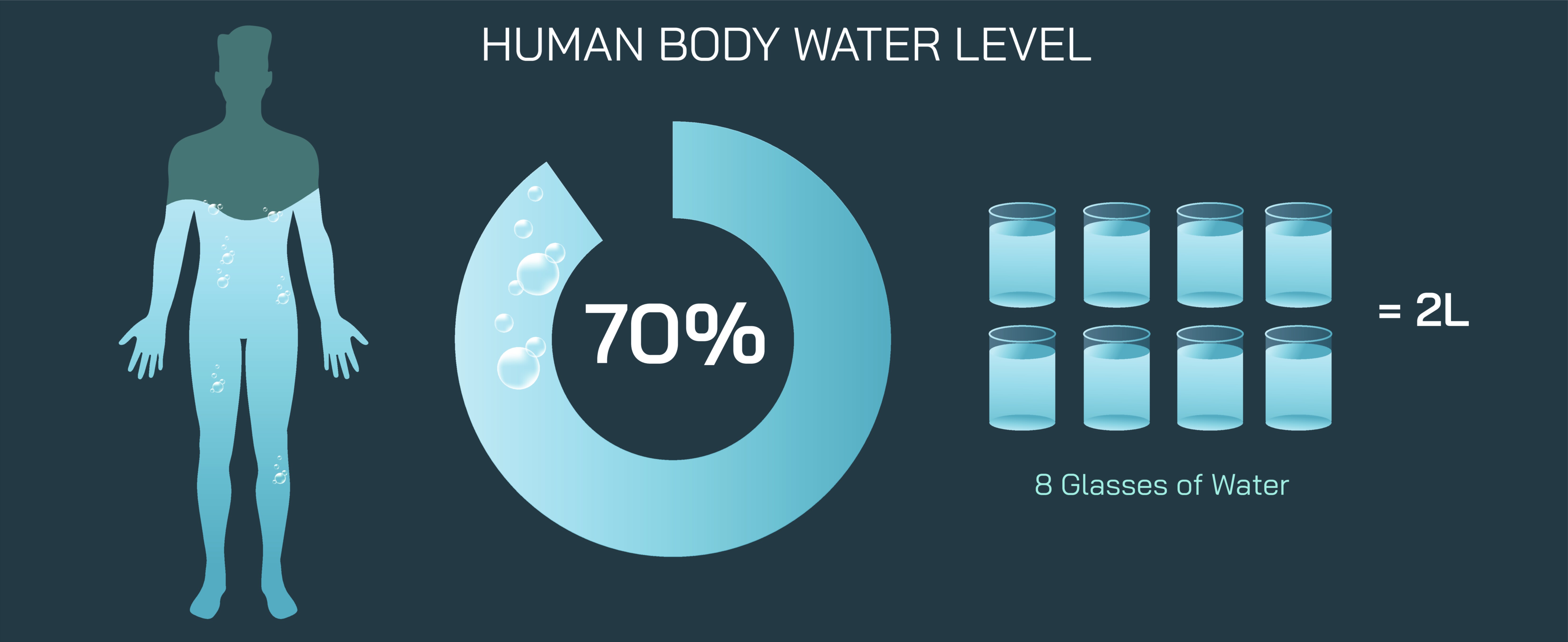
Human Body Water Level Vector Illustration Showing Percentage Distribution in Cells, Tissues, Blood, and Organs for Health Education
Water is the most abundant component of the human body, playing a vital role in maintaining homeostasis, supporting cellular function, transporting nutrients, and regulating temperature. In adults, water comprises approximately 60% of total body weight, distributed across intracellular fluid (ICF), extracellular fluid (ECF), blood plasma, and interstitial spaces. A vector illustration depicting human body water levels typically integrates percentage distribution, compartmental breakdown, and organ-specific water content, providing a clear, educational visualization of how water supports bodily functions. By combining labeled compartments, color-coded regions, and proportional graphics, such illustrations allow learners to understand both the quantity and functional significance of water in the human body.
At the center of the illustration is a human silhouette, representing total body composition. Color-coded segments divide water into major compartments: intracellular fluid (~40% of body weight), extracellular fluid (~20%), which includes interstitial fluid (~15%) and plasma (~5%). Arrows and labels indicate the flow of water between these compartments, visually emphasizing dynamic exchange and the principle that water distribution is crucial for homeostasis. Gradients or transparency effects may be used to highlight the proportion of water relative to body mass, helping learners immediately grasp quantitative relationships.
The intracellular compartment is illustrated as the largest portion of body water, contained within cells across all tissues and organs. Labels identify water-rich organs such as muscle (~75% water content), emphasizing the functional role of intracellular water in biochemical reactions, maintaining cell turgor, and enabling metabolic processes. Arrows may indicate intracellular water’s involvement in nutrient transport, enzymatic activity, and waste removal, connecting structural distribution to physiological importance. Color coding differentiates cytoplasmic fluid from extracellular fluid to clarify boundaries for educational purposes.
The extracellular fluid compartment is divided into interstitial fluid and blood plasma. Interstitial fluid surrounds cells in tissues, accounting for roughly 15% of body weight, while plasma constitutes approximately 5%. Vector diagrams often include magnified tissue insets, showing capillary networks, interstitial spaces, and fluid exchange via osmosis and filtration. Arrows indicate the movement of water from plasma to interstitial fluid and vice versa, emphasizing fluid balance and homeostatic regulation. Color differentiation helps learners distinguish plasma (often shown as red-tinted) from interstitial fluid (lighter blue or transparent), visually reinforcing compartmental distinctions.
Blood and cardiovascular components are illustrated to show how plasma water facilitates nutrient transport, gas exchange, and waste removal. Labels identify key elements such as plasma, red blood cells, and lymph, while arrows indicate direction of blood flow and water movement, linking circulation to systemic hydration and organ function. Insets may show kidneys and their role in water regulation, emphasizing physiological processes such as filtration, reabsorption, and urine production.
Organ-specific water content is highlighted to illustrate variability among tissues. For example, brain and kidneys (~73% water), lungs (~83%), liver (~70%), and adipose tissue (~10–20%) are labeled with proportional indicators or percentage overlays. Arrows may point to these organs from the central body silhouette, showing how hydration affects organ function and overall health. Color-coded gradients can reflect higher or lower water content, providing immediate visual cues about tissue composition and hydration needs.
Vector illustrations often include dynamic arrows and flow indicators, showing water exchange between intracellular and extracellular compartments. Labels may indicate osmosis, diffusion, and vascular transport, linking anatomical distribution to cellular and physiological mechanisms. Magnified panels may depict cell membranes with water movement, capillaries with plasma exchange, and tissue matrices, reinforcing functional understanding.
Additional features in educational illustrations include overall percentage breakdown, such as a pie chart or stacked bar, showing intracellular fluid, interstitial fluid, and plasma proportion. Legends clarify each compartment, providing learners with both quantitative and visual reference points. Comparative panels may also demonstrate hydrated versus dehydrated states, highlighting the consequences of fluid imbalance on organs, cells, and circulation.
By combining human silhouette, intracellular and extracellular compartments, organ-specific content, blood plasma, interstitial spaces, flow arrows, and percentage overlays, a human body water level vector illustration provides a comprehensive understanding of fluid distribution. Color coding, labeled compartments, and directional arrows clarify quantitative and functional aspects of hydration, making complex physiological concepts visually accessible.
Ultimately, a vector illustration of human body water distribution demonstrates the importance of water for cellular, tissue, blood, and organ function, linking anatomical distribution with physiological roles. Through labeled compartments, proportional shading, flow indicators, and organ-specific percentages, the diagram transforms abstract scientific data into an educational, visually engaging, and intuitive tool for health education, medical study, and wellness awareness.