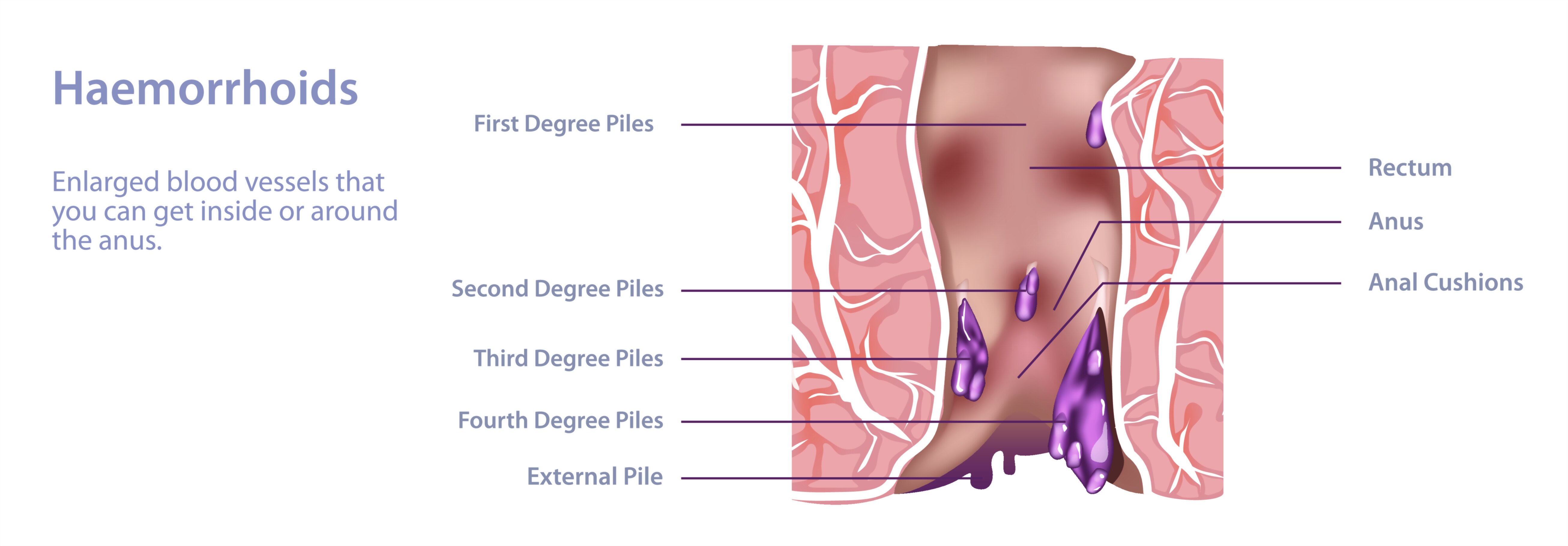
Hemorrhoids Vector Illustration Showing Swollen Veins in Rectum and Anus with Internal and External Types
Hemorrhoids, commonly known as piles, are a prevalent medical condition characterized by swollen, inflamed veins in the rectum and anus, resulting from increased pressure on the lower rectal blood vessels. They can cause discomfort, pain, itching, and bleeding during defecation. A vector illustration of hemorrhoids typically depicts the anatomical location, vascular structures, and differentiation between internal and external hemorrhoids, providing a detailed visual explanation for both patients and medical professionals. By combining anatomical accuracy with clear labeling and cross-sectional views, such illustrations enhance understanding of the condition, its causes, symptoms, and clinical relevance.
The illustration generally starts with the rectum and anal canal anatomy, showing the distal portion of the large intestine and the surrounding muscular structures, including the internal and external anal sphincters. The rectal wall contains a network of veins, known as hemorrhoidal plexuses, which are responsible for normal venous drainage but can become engorged under certain conditions. Vector diagrams highlight these veins with color coding—typically red or purple—to indicate inflammation and swelling. Cross-sectional views may show both the rectal lumen and the surrounding vascular structures, clarifying the location of internal and external hemorrhoids relative to the sphincter muscles and anal margin.
Internal hemorrhoids are located inside the rectum and are usually covered by mucosa. They are not typically visible from the outside and are often painless due to their location above the dentate line, where visceral nerves predominate. In vector illustrations, internal hemorrhoids are shown as engorged veins bulging into the rectal lumen. Labels may indicate stages of prolapse, ranging from small, non-protruding swellings to larger hemorrhoids that can protrude outside the anus during straining but retract spontaneously or manually. Arrows or shading can indicate the direction of venous expansion and the pressure exerted by straining or constipation, emphasizing the physiological mechanism behind their formation.
External hemorrhoids, by contrast, develop under the skin around the anus and are covered by sensitive tissue rich in somatic nerves, making them more painful when inflamed or thrombosed. In vector diagrams, external hemorrhoids are depicted as swollen, purplish nodules at the anal margin, sometimes with surrounding irritation or inflammation. Arrows may indicate venous congestion or thrombus formation, and color variations can illustrate severity and risk of complications. The diagram may also show how external hemorrhoids can become thrombosed, forming firm, painful lumps, helping viewers understand the clinical symptoms and the need for treatment in severe cases.
A comprehensive vector illustration often includes cross-sectional comparisons of internal and external hemorrhoids. These side-by-side views show the relative positions of swollen veins, anal sphincters, and rectal lining. Such representations help viewers distinguish between the types, clarify why pain perception differs, and demonstrate how treatment approaches may vary. For instance, internal hemorrhoids may be treated with dietary modification, topical ointments, or minimally invasive procedures, while external hemorrhoids may require conservative management or surgical intervention depending on severity.
Vector illustrations may also depict contributing factors, such as increased intra-abdominal pressure, constipation, prolonged sitting, pregnancy, and straining during bowel movements. Icons, arrows, or labels can indicate how these factors lead to venous dilation, congestion, and eventual formation of hemorrhoids. Some diagrams highlight venous anatomy, showing the connections between superior, middle, and inferior rectal veins, and how pressure imbalances can lead to localized swelling. By including this context, the illustration bridges anatomy with pathophysiology, enhancing comprehension of cause and effect.
Additional features in vector diagrams often include stages of hemorrhoid development, from mild swelling with minimal symptoms to advanced prolapsed or thrombosed hemorrhoids. Color gradients, arrows, or inset magnifications may show internal vascular changes, protrusion through the anal canal, and surrounding tissue inflammation. These elements allow learners to visualize progression and understand the correlation between anatomical changes and clinical symptoms.
Practical illustrations may also depict treatment strategies or preventive measures, such as increasing fiber intake, maintaining hydration, sitz baths, or procedural interventions like rubber band ligation, sclerotherapy, or surgical excision. While not always the primary focus, these elements help connect anatomical understanding with medical management, providing a comprehensive educational framework.
By integrating rectal and anal anatomy, venous structures, internal and external hemorrhoid locations, stages of swelling, and contributing factors, a hemorrhoid vector illustration offers a complete visual representation of the condition. It clarifies the distinction between internal and external types, demonstrates the mechanism of formation, and links anatomical changes to symptoms and treatment considerations. Color coding, cross-sectional views, labeled structures, and arrows representing pressure or venous expansion enhance clarity, making complex anatomy and pathology accessible.
Ultimately, a vector illustration of hemorrhoids communicates both the structural and functional aspects of swollen veins in the rectum and anus. It provides a visual understanding of how internal and external hemorrhoids develop, why symptoms vary, and how anatomical and physiological factors interact. By combining anatomical accuracy, clinical relevance, and illustrative clarity, the diagram serves as an essential educational tool for students, healthcare providers, and patients seeking to understand hemorrhoid formation, presentation, and management.