Enlarged Spleen Vector Illustration Showing Spleen Anatomy, Causes, and Effects on Blood Filtration
The spleen is a vital organ in the human lymphatic and circulatory systems, responsible for blood filtration, immune response, and red blood cell recycling. When the spleen becomes abnormally large, a condition known as splenomegaly, it can significantly affect the body’s ability to manage blood cells and fight infections. A vector illustration depicting an enlarged spleen typically includes the spleen’s anatomical structure, nearby organs, blood vessels, and visual markers indicating its increased size. Such diagrams also often incorporate information on the underlying causes of enlargement and the physiological effects on the circulatory system, making the illustration both educational and clinically informative.
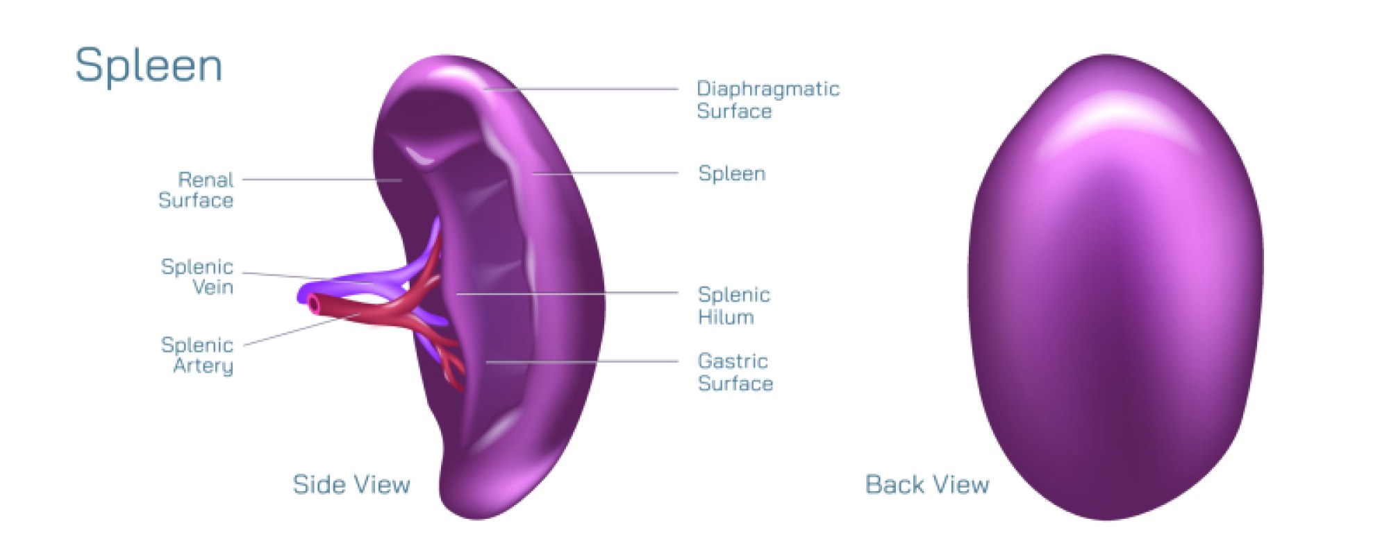
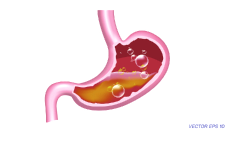
At the anatomical level, the spleen is located in the upper left quadrant of the abdomen, just beneath the diaphragm and adjacent to the stomach, kidney, and pancreas. In a vector illustration, the spleen is typically shown as a soft, oval organ with a rich vascular supply, highlighting the splenic artery and splenic vein that carry blood to and from the organ. The outer capsule of the spleen and internal structures, including the red pulp and white pulp, may be represented in cutaway form. The red pulp is responsible for filtering old or damaged red blood cells, while the white pulp contains lymphoid tissue that produces white blood cells and antibodies, contributing to immune defense. Illustrating these components helps viewers understand the dual functions of blood filtration and immune response within the spleen.
In cases of splenomegaly, the spleen enlarges beyond its normal size, which can be depicted in a vector diagram by increasing its relative size compared to adjacent organs. This enlargement may cause displacement or compression of nearby structures, which can be highlighted in the illustration with arrows or shading. An enlarged spleen is often palpable below the left rib cage and may be associated with abdominal discomfort, early satiety due to stomach compression, and tenderness. Visual representations may include color-coded areas to show increased blood pooling or congestion within the spleen, illustrating the mechanical impact of organ enlargement on circulatory function.
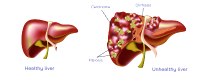
Causes of an enlarged spleen are diverse and can be effectively illustrated in a vector diagram through symbolic icons, labels, or flow charts. Common causes include infections, such as mononucleosis or bacterial endocarditis; liver diseases, such as cirrhosis causing portal hypertension; hematologic disorders, including hemolytic anemia, sickle cell disease, or leukemia; and inflammatory or autoimmune conditions, such as lupus or rheumatoid arthritis. Vector illustrations often depict these causes as linked to the spleen with arrows, helping viewers understand the systemic relationships between disease processes and organ enlargement. Additionally, congenital or genetic conditions may be highlighted to show hereditary factors contributing to spleen pathology.
The effects of an enlarged spleen on blood filtration and immune function are central to the diagram’s educational purpose. As the spleen enlarges, it can become hyperactive, a condition known as hypersplenism, leading to excessive sequestration of red blood cells, white blood cells, and platelets. This results in anemia, leukopenia, or thrombocytopenia, which can be depicted in the vector illustration with symbols representing reduced blood cell counts and arrows indicating sequestration or destruction within the enlarged organ. The diagram may also highlight the spleen’s impact on the immune system, showing that while the spleen continues to produce antibodies and filter pathogens, its overactivity can compromise blood cell levels, leading to increased susceptibility to infections and bleeding disorders.
Vector illustrations may further show the relationship of the spleen with the liver and portal circulation, as portal hypertension can contribute to splenomegaly. The splenic vein, flowing into the portal vein, may be labeled, and enlarged spleens can be visually connected to congestion in this vascular network. This representation helps viewers understand how systemic circulation and pressure changes contribute to the organ’s enlargement and associated complications.
Clinical features and potential complications can also be integrated into the illustration. These may include abdominal fullness, left upper quadrant pain, fatigue from anemia, and increased risk of infections. Vector arrows, labels, or color-coded markers may be used to show these systemic effects, linking the anatomical enlargement of the spleen to the broader physiological and clinical consequences. Additional features may include visual comparisons of a normal versus enlarged spleen, showing size differences and how the enlargement impacts neighboring organs and structures.
By combining anatomical accuracy, vascular detail, and pathological context, the vector illustration of an enlarged spleen serves multiple educational purposes. It clearly demonstrates the location, size, and internal structures of the spleen, while also visually explaining the causes of enlargement and its systemic effects on blood filtration and immunity. Such diagrams are invaluable in medical education, patient awareness, and clinical assessment, as they bridge the gap between abstract concepts and tangible anatomical and physiological understanding.
Ultimately, a vector illustration of an enlarged spleen emphasizes both the structural and functional consequences of splenomegaly. It conveys the dual role of the spleen as a blood filter and immune organ, how these functions are affected by enlargement, and the clinical implications for blood cell counts and systemic health. By integrating anatomy, pathology, and physiology into a single visual representation, the diagram provides a comprehensive educational tool that elucidates the complex interplay between organ size, function, and systemic disease, allowing learners to grasp the significance of spleen enlargement in a clear and visually intuitive manner.