Human Hand Bones Anatomy Vector Illustration Showing Carpals, Metacarpals, and Phalanges with Labeled Structure
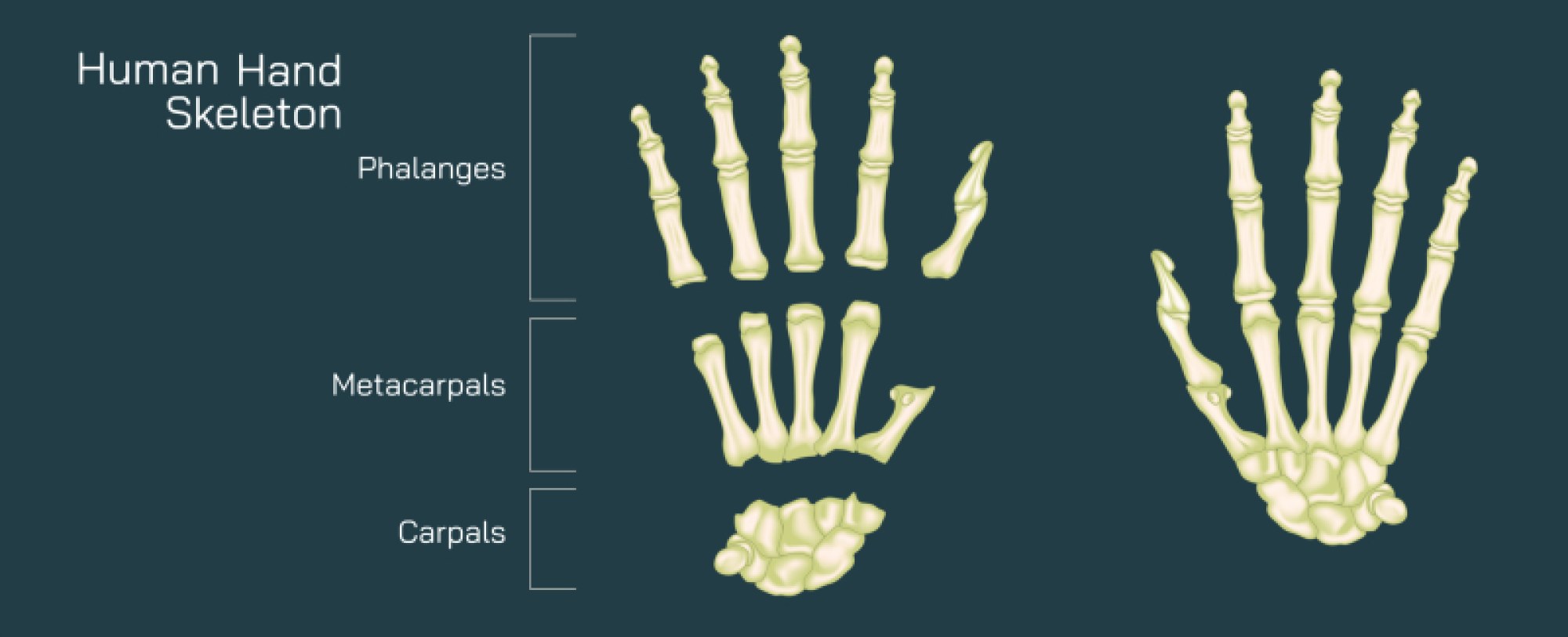
The human hand is a marvel of anatomical engineering, combining strength, flexibility, and fine motor control. Its skeletal framework, consisting of carpals, metacarpals, and phalanges, forms the foundation for a wide range of movements, from powerful grips to delicate manipulations. A vector illustration of the human hand bones typically highlights these skeletal components with clear labels, anatomical divisions, and joint articulations, providing a detailed, educational view of the hand’s structure. Such diagrams are essential for medical education, physiotherapy, orthopedics, and biomechanics, allowing learners to understand the arrangement, interconnection, and function of each bone in the hand.
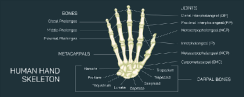
At the base of the hand are the carpal bones, eight small, irregularly shaped bones arranged in two rows within the wrist. These bones create a strong yet flexible platform for hand motion and bear the forces transmitted from the forearm to the palm. The proximal row, closest to the forearm, consists of the scaphoid, lunate, triquetrum, and pisiform, while the distal row, adjacent to the palm, includes the trapezium, trapezoid, capitate, and hamate. In vector illustrations, each carpal bone is clearly labeled, often with color coding to differentiate the proximal and distal rows. Arrows or callouts may indicate articulations with adjacent bones and joints, showing how the carpal cluster facilitates wrist flexion, extension, abduction, and adduction. Highlighting the anatomical arrangement helps viewers understand how these bones form a stable base for the metacarpals while permitting multidirectional movement.
Extending distally from the carpals are the metacarpal bones, five long bones that form the skeletal structure of the palm. Each metacarpal has a base, shaft, and head, with the bases articulating with the distal carpal bones and the heads forming the knuckles at the metacarpophalangeal (MCP) joints. Vector diagrams typically number the metacarpals from one (thumb side) to five (little finger side), and may use different colors or labels to emphasize their position and function. The first metacarpal is shorter and more mobile than the others, enabling the thumb’s unique opposition movement, which is crucial for gripping and manipulating objects. Arrows may illustrate the movement range at each MCP joint, showing flexion, extension, abduction, and adduction capabilities.
The phalanges form the skeleton of the fingers and thumb. Each finger has three phalanges: proximal, middle, and distal, while the thumb has only two: proximal and distal. Vector illustrations often depict the alignment of phalanges in relation to the metacarpals, highlighting the interphalangeal (IP) joints, which allow bending and straightening of the digits. Proximal interphalangeal (PIP) and distal interphalangeal (DIP) joints in the fingers, along with the interphalangeal joint in the thumb, are commonly labeled and may include arrows demonstrating the range of motion. Color-coded phalanges or distinct shading may further enhance clarity, allowing viewers to differentiate between the three segments of the fingers and the two of the thumb.
A comprehensive vector illustration also emphasizes joint articulation and skeletal interconnections. The carpometacarpal (CMC) joints connect the metacarpals to the distal row of carpal bones, while the metacarpophalangeal (MCP) joints link metacarpals to proximal phalanges. Interphalangeal joints connect the phalanges themselves. The thumb’s saddle-shaped CMC joint is often highlighted to show its contribution to opposition and precise hand function. Arrows, lines, or gradient shading in the diagram may illustrate the directions of movement, demonstrating how the skeletal framework facilitates both powerful gripping motions and delicate manipulations.
Vector illustrations frequently include multiple perspectives—dorsal, palmar, and lateral views—to provide a three-dimensional understanding of the hand skeleton. Labels identify each bone, joint, and key anatomical landmark, ensuring that all components are clearly understood in relation to one another. Magnified insets may focus on smaller, clinically significant bones such as the scaphoid or lunate, which are prone to fractures or instability. Color coding, numbering, and directional arrows further enhance comprehension, making the diagram suitable for detailed study and clinical reference.
Additional elements in the illustration may depict functional and clinical context. For instance, the alignment of metacarpals and phalanges may be shown during gripping or finger flexion, illustrating how the skeletal arrangement supports hand function. Common fracture sites, such as the scaphoid or distal phalanges, may be indicated, providing practical relevance for medical education and orthopedic practice. The illustration can also highlight tendon attachment points or ligament connections to explain how skeletal structure interacts with the muscular and connective tissue systems for coordinated movement.
By combining carpals, metacarpals, and phalanges with joint structures, labels, and functional annotations, a vector illustration of the human hand skeleton provides a comprehensive view of the hand’s anatomy. It shows how bones articulate, transmit forces, and allow a range of movements essential for daily tasks, sports, and skilled manual work. The diagram clarifies spatial relationships, structural hierarchy, and the mechanical principles underlying hand function.
Ultimately, a hand skeleton vector illustration demonstrates the complex interplay between skeletal elements, joint articulations, and movement potential. It emphasizes how the carpals provide stability and flexibility, metacarpals support the palm and knuckles, and phalanges enable precise finger motions. Through detailed labeling, multi-view representation, and functional depiction, the diagram transforms the intricate anatomy of the hand into a visually intuitive educational tool, enhancing comprehension of both structure and practical biomechanics.