Femur Bone Showing Structure, Shape, and Detailed Anatomy of the Longest Human Skeletal Bone
The femur is the most powerful architectural component of the human skeleton, and its anatomy embodies a unique fusion of structural strength, biomechanical efficiency, and biological function. Extending from the hip to the knee, this bone forms the backbone of the lower limb and makes upright posture, walking, and running possible by resisting immense vertical and lateral loads with remarkable resilience. A detailed visualization of the femur—its shape, anatomical landmarks, and internal composition—reveals how every curve, surface, ridge, and cavity is purpose-built to withstand weight-bearing forces while enabling fluid, coordinated movement through the hip and knee. Understanding the femur in detail is not simply memorizing its parts; it means appreciating how the skeletal system evolved to make powerful motion safe, stable, and energy-efficient.
At the uppermost end lies the femoral head, a spherical structure that articulates with the pelvis at the acetabulum to form the hip joint. This ball-and-socket design provides the broadest range of motion of any lower limb joint, permitting flexion, extension, abduction, adduction, circumduction, and rotation without compromising stability during standing or locomotion. Thick articular cartilage coats the femoral head, distributing weight and minimizing friction as the hip moves. Supporting the spherical head is the femoral neck, a slim yet strong bridge positioned at an angle that optimizes leverage and allows for wide hip mobility while aligning the head into the pelvic socket. Although efficient for movement, this geometry makes the neck a clinically significant site for fractures, especially in individuals with compromised bone density, where rotational and compressive forces converge.
Projecting from the neck are two prominent outgrowths—the greater trochanter and lesser trochanter—which serve as major muscle attachment sites. The greater trochanter anchors the powerful gluteus medius and gluteus minimus muscles, which stabilize the pelvis during single-leg stance. The lesser trochanter serves as the insertion point for the iliopsoas, the primary hip flexor responsible for raising the thigh during walking and running. These trochanters convert muscle contractions into directional force, reinforcing that the femur is as much a lever system as it is a support column. In detailed anatomical visuals, the trochanters stand out as bony landmarks where mechanical energy is transferred from muscle to motion.
Below these projections stretches the shaft of the femur, a long cylindrical segment with a subtle anterior bow. This curvature is not accidental—it helps the bone dissipate compressive stress during standing and impact, preventing fractures under heavy loads. The shaft’s walls are composed of compact cortical bone, a dense matrix that resists bending and torsion. Internally, the bone transitions to trabecular (spongy) bone, which is arranged in lattice-like patterns that reflect stress distribution across the limb. A prominent ridge along the posterior shaft called the linea aspera serves as a linear anchor for muscles such as the adductor group and the vastus medialis. This feature is crucial in gait mechanics because it balances muscular pull on the femur and supports coordinated knee flexion and hip stabilization.
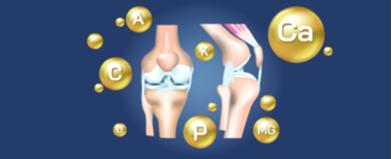
Approaching the lower extremity of the bone, the femur expands into two rounded structures—the medial and lateral condyles—which articulate with the tibia and form the weight-bearing portion of the knee joint. These smooth condylar surfaces are separated posteriorly by the intercondylar fossa, which houses the cruciate ligaments that prevent anterior and posterior shifting of the tibia. On the anterior aspect, the patellar surface creates a track for the patella during knee extension and flexion, enabling quadriceps efficiency by enhancing leverage. The shape of the condyles ensures even weight distribution and stable motion across the knee throughout activities such as climbing stairs, squatting, running, and sudden directional changes.
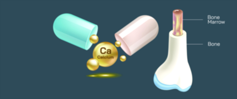
Internally, the femur houses the medullary cavity, a long tubular space containing bone marrow, making the femur not only a structural pillar but a metabolic and hematologic organ. In children and young adults, the cavity is filled with red marrow, responsible for the production of blood cells—red blood cells, white blood cells, and platelets. As the skeleton matures, a large portion of this marrow transitions to yellow marrow, which stores lipids that serve as energy reserves and can convert back to red marrow under intense physiological demand. The presence of marrow highlights that the femur is as vital to immune and oxygen transport systems as it is to biomechanical strength.
The functions of the femur emerge from the integration of its anatomical features. Structurally, it bears body weight, transferring it from the pelvis to the knee while absorbing shock during every step. Biomechanically, it acts as a lever for major muscles of the hip and thigh, enabling propulsion and maintaining balance. Architecturally, its external contours and internal composition protect arteries, veins, and nerves running alongside it. Physiologically, its marrow sustains blood production and mineral storage, allowing the femur to act as a reserve for calcium and phosphorus needed for metabolic processes. This multidimensional role demonstrates the femur’s importance not just for mobility but for life-supporting biological stability.
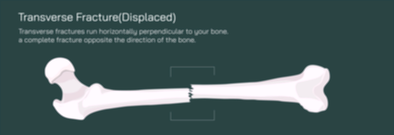
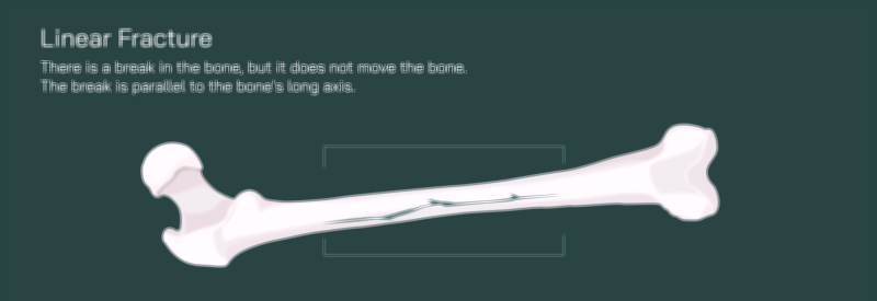
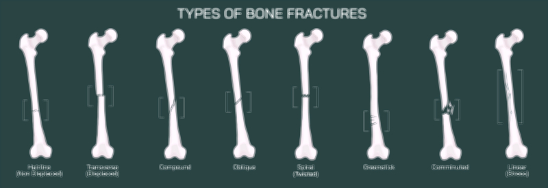
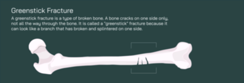
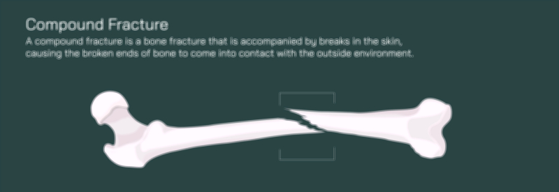
The relationships between regions explain why injuries in different locations behave differently. A fracture at the femoral neck may impair hip rotation and require surgical stabilization; an injury to the shaft affects load transmission and muscle attachment points; a fracture through the condyles impacts knee stability and gait. Likewise, degenerative changes in the hip joint can stem from narrowing cartilage on the femoral head, while knee pain or patellar tracking issues can relate to the patellar surface or muscular imbalance along the linea aspera. These clinical variations are easier to understand when the femur is seen as a system in which every surface and ridge influences movement, posture, and weight distribution.
When represented in detailed anatomical graphics, the femur becomes more than a single bone—it becomes a map of how humans stand upright, maintain balance, and perform complex movement with efficiency and power. It reveals the logic behind muscle connections, the purpose of joint engineering, the adaptability of bone under stress, and the biological significance of the marrow hidden deep within. Examining the femur closely encourages an understanding of the body as a coordinated mechanism rather than a series of isolated parts.
Ultimately, the femur’s structure and shape are a reflection of its purpose: a long, robust lever that balances durability with flexibility, houses critical physiological tissue, and enables the smooth integration of motion from hip to knee. Through its dense cortical walls, curved shaft, precisely formed joint surfaces, muscle attachment prominences, and living marrow core, the femur supports human mobility from childhood through old age. A detailed anatomical view not only deepens appreciation for skeletal design but also illuminates how vital this single bone is to independence, posture, movement, and daily life.