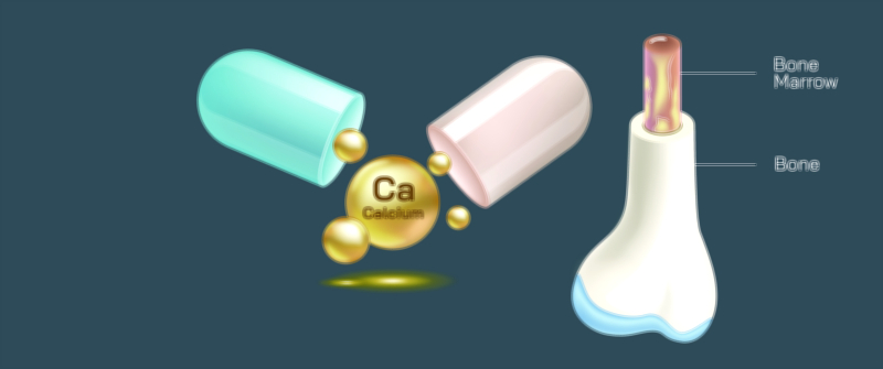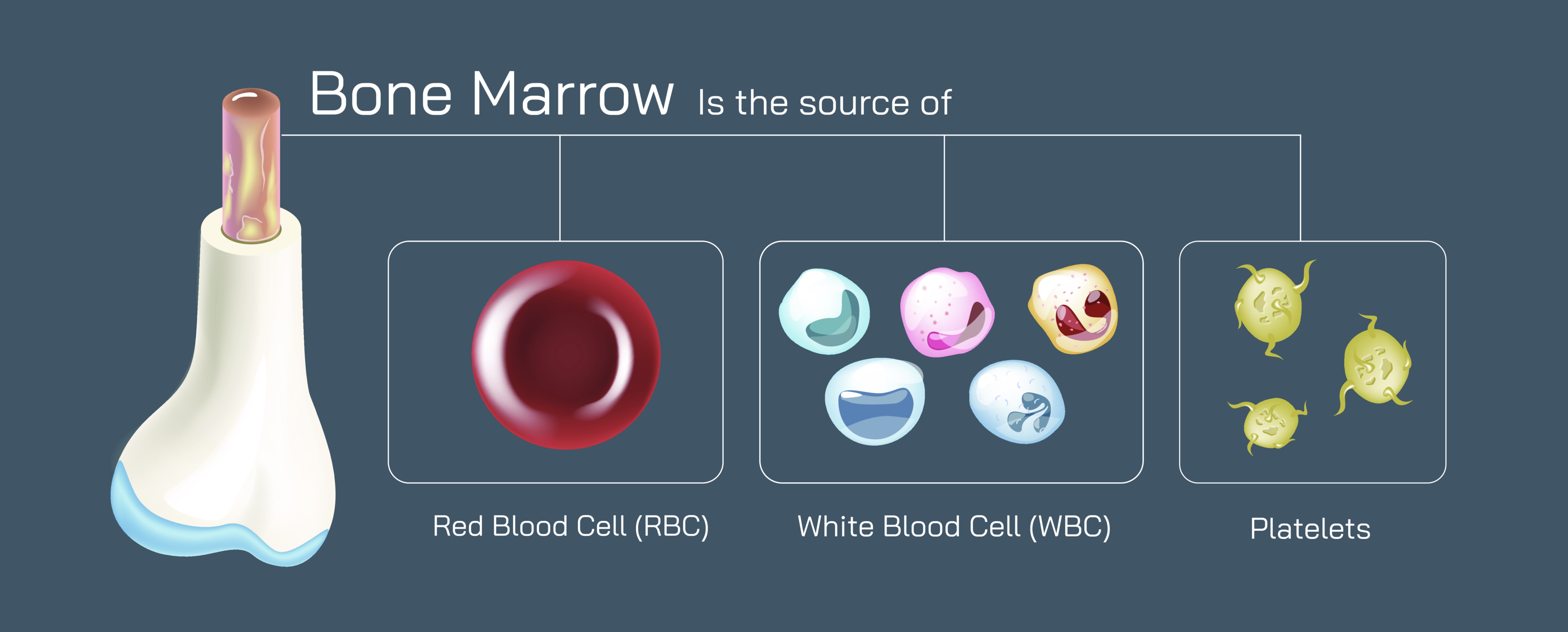
Bone Marrow Components Vector Illustration Showing Red and Yellow Marrow with Blood Cell Formation
Bone marrow is one of the most critical yet least visible tissues within the human body, and understanding its structure is essential for grasping how blood is produced and how immunity and oxygen delivery are sustained throughout life. A vector illustration showing the two major components of bone marrow—red marrow and yellow marrow—along with their functions in blood cell formation provides a powerful learning tool for anatomy, physiology, hematology, and medical education. Bone marrow lies deep inside bones, filling the spaces within the spongy trabecular network and functioning as the body’s core blood cell manufacturing system. Although the skeleton is often associated with structure and strength, the marrow inside it plays a metabolic role just as important as the heart, lungs, or brain. By visually differentiating red and yellow marrow and mapping out the cells each one produces, a medical illustration brings to life a process that is silently ongoing every second.
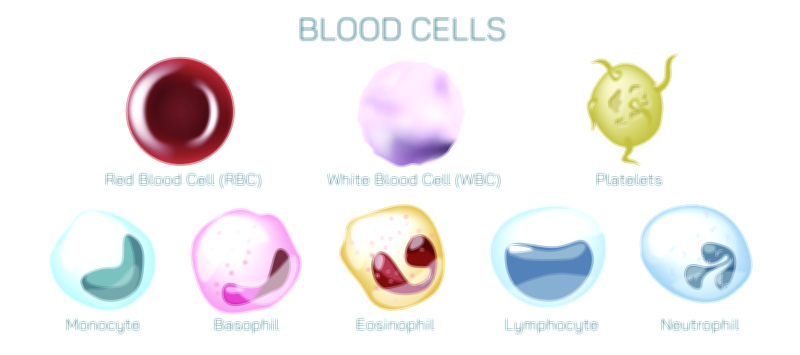
Red bone marrow is the body's primary hematopoietic tissue—the tissue responsible for building blood. It produces red blood cells (RBCs), white blood cells (WBCs), and platelets, a collective process known as hematopoiesis. When represented visually, red marrow is typically shown with vibrant shades symbolizing activity and density. A cross-sectional vector illustration of a bone will often highlight red marrow occupying the interior spaces of flat bones and the ends of long bones, where active trabecular bone provides a supportive, highly vascular environment. The reason for its deep red coloration lies in the high concentration of developing RBCs and iron-rich hemoglobin, the pigment that ultimately transports oxygen throughout the body. Red marrow also houses multipotent hematopoietic stem cells, the parent cells from which all blood cells descend. These stem cells differentiate along diverse pathways: RBCs that carry oxygen, WBCs that protect against pathogens, and platelets that enable clotting and wound healing. A well-designed illustration showing branching differentiation pathways helps learners visualize how one population of stem cells can develop into so many vital cellular types.
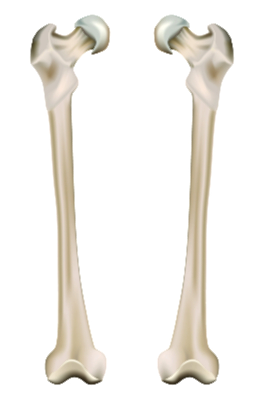
Yellow marrow, in contrast, is composed predominantly of adipocytes (fat cells) and serves as the body’s marrow-based energy reservoir. While red marrow is associated with intense metabolic activity, yellow marrow represents a nutrient storage center that can be activated under stress. A vector diagram usually depicts yellow marrow in softer golden or pale hues to represent its lipid richness. Yellow marrow is primarily located in the central shafts (medullary cavities) of long bones, such as the femur, tibia, and humerus. Even though yellow marrow’s primary composition is fat, it retains hematopoietic potential: in cases of severe blood loss or illness that increases blood cell demand, the body can convert yellow marrow back into red marrow to expand blood cell production. Illustrating this reversible transformation allows learners to understand marrow not as static tissue but as adaptable biological machinery that adjusts to the body's needs across the lifespan.
The distribution of red and yellow marrow changes dramatically with age, and this developmental progression is especially important to convey visually. In newborns and young children, nearly all marrow is red because the body requires continuous production of new blood cells to support growth. As a person ages, red marrow gradually converts to yellow marrow, especially in long bone shafts, while remaining red and active in flat bones such as the sternum, pelvis, ribs, and vertebrae. By adulthood, about half of total bone marrow is red and half is yellow, balancing blood cell production with energy storage. Educational images that show this distribution across childhood, adulthood, and older age make the connection between marrow composition and health easier to understand. They also reinforce why bone marrow biopsies are commonly taken from pelvic bones—they contain red marrow throughout life.
Each type of marrow plays distinct physiological roles, and a vector diagram that shows the cellular workflow inside the bone helps viewers connect structure with function. Red marrow constantly forms RBCs that circulate oxygen, WBCs that fight infection, and platelets that close wounds. Yellow marrow stores fats that support metabolism during shortages, injury, or illness. Together, they ensure that bones remain both structural supports and life-sustaining tissues. Illustrations frequently include sinusoidal capillaries within red marrow to demonstrate how newly formed blood cells enter the bloodstream. These vessels possess porous walls that allow developed cells to pass from the marrow into circulation—another detail that medical visuals clarify far more effectively than text alone.
A marrow-focused vector illustration also provides a gateway to recognizing disease processes. Disorders such as leukemia, lymphoma, aplastic anemia, multiple myeloma, and myelodysplastic syndromes originate in or affect the marrow and disrupt blood cell production. Visual representations help explain how malignant or dysfunctional cells can replace healthy hematopoietic cells and interfere with normal development. Likewise, conditions such as anemia, infection susceptibility, and clotting problems make more sense when viewers recognize that marrow health determines the availability of blood cells. Even in orthopedic injuries, such as fractures, marrow can play a role by releasing fat into circulation or rapidly increasing blood cell output to support healing. In clinical settings, understanding marrow architecture makes procedures such as bone marrow biopsies, transplants, and stem cell therapies easier to conceptualize for patients and students.
The immune dimension of marrow is also notable. Red marrow contains early precursors of lymphoid cells, which then migrate to the thymus or lymph nodes to mature into T cells and B cells—critical defenders in the adaptive immune system. A vector illustration that links marrow to immune organs (thymus, lymph nodes, spleen) clarifies that immunity does not originate solely from lymphatic structures but begins deep within bones.
From a nutritional and lifestyle perspective, the health of marrow depends on adequate intake of iron, vitamin B12, folate, protein, and other micronutrients; maintaining sufficient physical activity; and avoiding toxins such as smoking and alcohol excess that interfere with blood formation. While such factors may not be the main focus of the vector illustration, many educational layouts include visual cues around the bone to show the environmental influences that support or weaken marrow activity.
In its full purpose, a bone marrow components vector illustration showing red and yellow marrow with blood cell formation does far more than display anatomy. It reveals that bones are not passive beams of support—they house the machinery that oxygenates tissues, powers the immune system, and ensures the body can heal from injury. By visually tracing blood cells from their earliest stem cell form in red marrow to their full function in circulation, and by contrasting the vibrant cellular activity of red marrow with the energy-rich composition of yellow marrow, the illustration brings together structure, biochemistry, physiology, and clinical relevance. It becomes a complete educational portrait of how marrow sustains life from within the skeleton, turning complex hematologic processes into a clear narrative that students, clinicians, researchers, and patients can instantly understand.