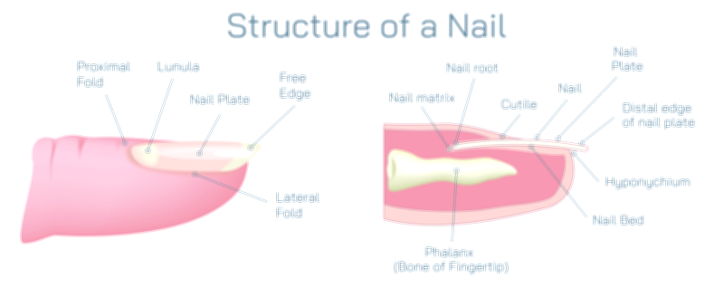
Finger Nail Showing Nail Plate, Cuticle, Matrix, Lunula, and Nail Bed Anatomy in the Human Hand
A fingernail may look like a simple hard surface resting on the end of the finger, but beneath its polished exterior lies a coordinated biological design built for protection, sensation, precision, and continuous growth. A vector illustration that highlights the nail plate, cuticle, matrix, lunula, and nail bed allows us to see the hidden layers of tissue that make everyday finger movement and fine motor tasks possible. Each component of the nail unit has a specific structure and function, and together they form a living system that protects the fingertip, enhances touch, and constantly renews itself through the growth of keratin.
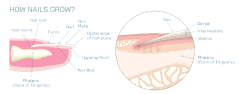
At the outermost surface is the nail plate, the visible hard portion of the nail. It is composed of multiple compact layers of keratinized cells tightly pressed together, creating a translucent shield that is both strong and flexible. Its curvature matches the contour of the fingertip, giving it the ability to withstand pressure and friction while allowing fingertips to grip, scratch, peel, pinch, and manipulate objects with remarkable precision. Because the nail plate is semi-transparent, the color of the underlying vascular tissue shows through, which is why healthy nails appear pinkish. The plate itself does not grow from the free edge — it continually advances outward from the base of the nail as new cells are formed underneath.
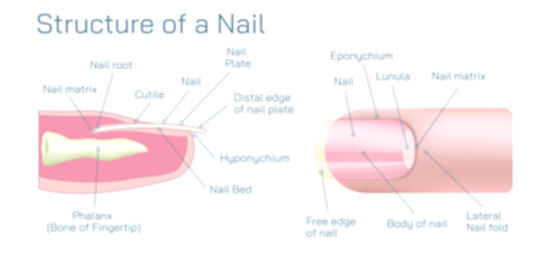
At the base of the nail plate lies the cuticle, or eponychium, a small but crucial structure that acts as a natural protective seal. The cuticle is a thin edge of skin that adheres tightly to the surface of the nail where it emerges from under the proximal nail fold. This seal prevents water, dirt, and microorganisms from sliding beneath the nail and reaching vulnerable tissues such as the matrix and nail bed. A damaged or aggressively trimmed cuticle weakens this barrier, making infections and inflammation more likely. Despite its modest size, the cuticle is essential for the long-term stability and health of the nail, protecting the area where new nail tissue begins its development.
The origin of nail growth is found beneath the skin in the nail matrix, the most biologically active part of the nail system. The matrix contains rapidly dividing cells that create new nail plate material. These newly formed cells begin soft and living but gradually harden through keratinization, flattening into the rigid keratin that ultimately becomes the nail plate. Because new nail cells are continually added at the base, older cells are pushed forward toward the tip of the finger, creating the steady growth that lengthens the nail over time. The shape, thickness, and smoothness of the nail depend on the condition of the matrix, which is why trauma or disease affecting this area can permanently alter nail appearance.
The lunula, the pale crescent-shaped area sometimes visible at the base of the nail plate, marks the frontmost portion of the matrix. Its lighter color occurs because the keratinization process within this zone has begun but is not fully complete, making the semi-formed nail cells less transparent to the pink coloration of the nail bed. The lunula acts as the bridge between deep matrix activity and the surface of the emerging nail plate. Its size and visibility vary between individuals and between fingers, but when present, it serves as a visible indicator of where nail formation transitions from soft to hardened keratin.
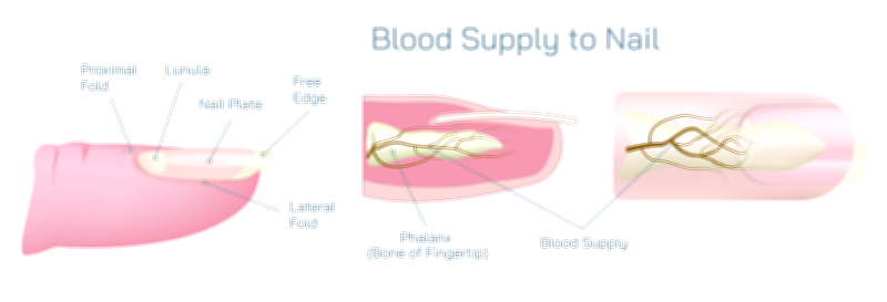
Beneath the nail plate runs the nail bed, the supportive and nourished layer that anchors the nail securely to the finger. It consists of specialized epithelium fused to the underside of the nail plate so closely that the two almost behave as a single unit. Capillary-rich tissue within the nail bed supplies nutrients and oxygen to the surrounding structures, reinforcing nail health and lending its vascular pink tone to the translucent plate above. Although the nail bed does not create the nail plate, it guides its smooth progression forward and stabilizes it until the plate reaches the fingertip’s free edge, where it eventually detaches from the bed.
Together, these anatomical components form a system balanced between strength and biological renewal. The nail protects the fingertip during everyday tasks, increases tactile sensitivity by providing a firm counter-surface for touch receptors beneath it, and acts as a tool for fine movements. The cuticle ensures a sterile pathway for growth, the matrix continuously manufactures new keratin cells, the lunula reflects the early visible stage of this keratinization process, and the nail bed anchors and protects the plate as it advances.
A complete illustration typically presents the nail in cross-section so that the visible structures on the surface and the hidden tissues underneath appear together, each labeled to reflect their role in nail function. When viewed this way, it becomes clear that a fingernail is not simply a cosmetic feature but a miniature biological mechanism — a protective barrier strengthened by keratin, a sensory support structure for the fingertip, and a continuously renewing tissue guided by the matrix and protected by the cuticle.
The anatomy of the fingernail demonstrates how the body integrates protection, growth, sensation, and precision in a small yet vital structure. Every component — the nail plate, cuticle, matrix, lunula, and nail bed — works in harmony to keep the fingertip powerful, sensitive, and resilient, allowing the hands to perform the delicate tasks that define human dexterity and touch.