Human Reproduction System Vector Illustration Showing Male and Female Reproductive Organs and Fertilization Process
The human reproductive system is a highly specialized biological system responsible for producing gametes, enabling fertilization, and supporting the development of offspring. A comprehensive vector illustration of the human reproductive system typically includes both male and female reproductive organs, alongside the fertilization process, providing a clear depiction of anatomy, physiology, and the intricate interaction between male and female reproductive mechanisms. Such illustrations serve as essential educational tools for students, healthcare professionals, and individuals seeking to understand the biological foundations of reproduction, including gamete production, fertilization, and early embryonic development.
The male reproductive system consists of organs that produce, store, and transport sperm cells while also synthesizing hormones such as testosterone, which regulates secondary sexual characteristics and reproductive function. A vector illustration of the male reproductive system usually highlights the testes, epididymis, vas deferens, seminal vesicles, prostate gland, urethra, and penis. The testes, located within the scrotum, are the primary male reproductive organs, producing sperm in seminiferous tubules and secreting testosterone. Vector diagrams often include a magnified inset showing seminiferous tubules with developing spermatozoa, emphasizing the process of spermatogenesis, which transforms germ cells into mature sperm over a period of approximately 64 days.
The epididymis, a coiled tube atop each testis, stores and matures sperm, preparing them for motility and fertilization capability. Arrows in a vector illustration may show the path of sperm from the testes through the epididymis and into the vas deferens, the muscular tube that transports sperm during ejaculation. The seminal vesicles contribute nutrient-rich fluid to sperm, providing fructose and other substances to support motility, while the prostate gland secretes alkaline fluid that neutralizes acidic vaginal conditions, enhancing sperm survival. The urethra serves as the conduit for semen during ejaculation and for urine excretion, while the penis delivers sperm into the female reproductive tract. Arrows and flow diagrams in vector illustrations typically trace the journey of sperm from production to deposition, providing a clear visualization of the male reproductive pathway.
The female reproductive system encompasses organs that produce eggs (ova), facilitate fertilization, and support embryonic and fetal development. A vector illustration of the female system commonly highlights the ovaries, fallopian tubes (oviducts), uterus, cervix, and vagina. The ovaries are paired glands that produce eggs and secrete hormones such as estrogen and progesterone. The illustration may include a magnified inset showing developing follicles within the ovary, illustrating oogenesis, the process by which primary oocytes mature into secondary oocytes ready for ovulation. The fallopian tubes extend from the ovaries to the uterus and serve as the site of fertilization; arrows in the illustration often indicate the movement of the egg toward the uterus, guided by ciliary action and muscular contractions.
The uterus is a muscular, pear-shaped organ that provides a nurturing environment for the developing embryo. The endometrium, the inner lining, thickens in response to hormonal signals during the menstrual cycle to prepare for implantation. The cervix forms the narrow lower segment of the uterus, connecting to the vagina, which serves as the passageway for sperm entry and the birth canal during delivery. In vector illustrations, the uterine structure is often cross-sectioned to show the endometrium, myometrium, and lumen, providing insight into the physiological environment encountered by the fertilized egg.
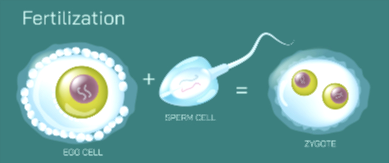
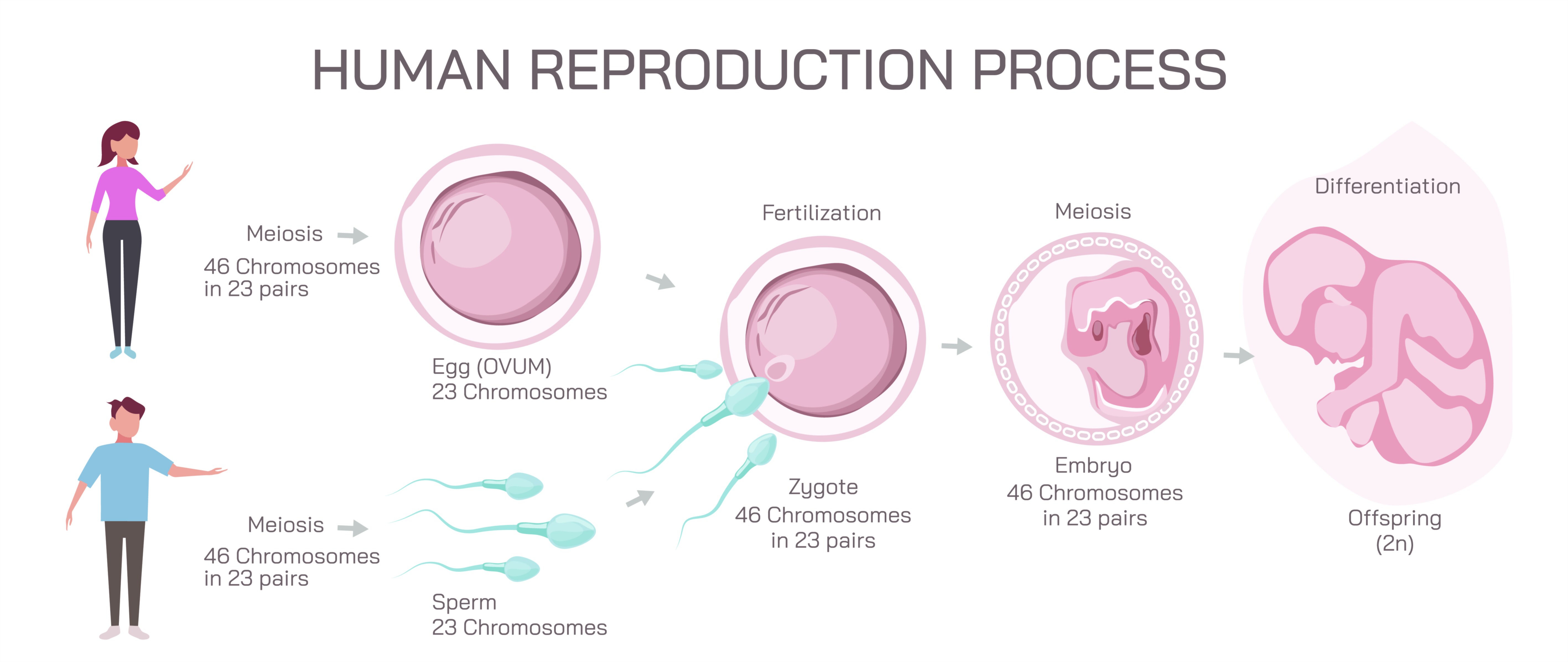
The fertilization process is a central feature of such illustrations. Sperm deposited in the vagina during intercourse traverse the cervix and uterus to reach the fallopian tube, where fertilization typically occurs. A vector diagram may depict a single sperm penetrating the ovum, representing the moment of zygote formation. Arrows may indicate the direction of sperm movement and the fusion of genetic material from both gametes, producing a diploid zygote. Following fertilization, the zygote begins cell division (cleavage) as it travels toward the uterus, eventually implanting in the prepared endometrium, initiating embryonic development. Illustrations may show this sequence with numbered steps or color-coded arrows to differentiate stages.
Vector diagrams may also emphasize hormonal regulation, showing how the hypothalamus-pituitary-ovarian axis controls ovulation, corpus luteum function, and endometrial preparation. Similarly, the hypothalamus-pituitary-testicular axis regulates sperm production, testosterone secretion, and reproductive behavior in males. Arrows and labeled pathways in the illustration help connect endocrine function to anatomical structures and reproductive outcomes.
Additional elements in the vector illustration may include comparative anatomy, highlighting the spatial relationship between male and female reproductive organs during copulation, and external features, such as the scrotum and vulva, for comprehensive anatomical context. Color-coded labels and magnified sections of ovaries, testes, and fallopian tubes enhance clarity, while arrows indicating gamete and embryo movement provide dynamic understanding of the reproductive process.
By integrating male and female anatomy with the fertilization process, a vector illustration of the human reproductive system provides a holistic view of reproduction. It demonstrates the production and maturation of gametes, the journey of sperm and ova, the mechanics of fertilization, and the initial stages of embryonic development. This visual representation links anatomy, physiology, and cellular processes, making it an essential educational tool for understanding human reproduction, sexual health, fertility, and developmental biology.
Ultimately, the vector illustration conveys the coordinated interplay of male and female reproductive systems. It emphasizes how anatomical structures, physiological mechanisms, and hormonal regulation work together to facilitate reproduction, illustrating the complexity, efficiency, and elegance of human reproductive biology. Through detailed labeling, color coding, arrows, and magnified sections, the diagram transforms abstract biological processes into an accessible and educational visual framework, suitable for medical, academic, and public health education.