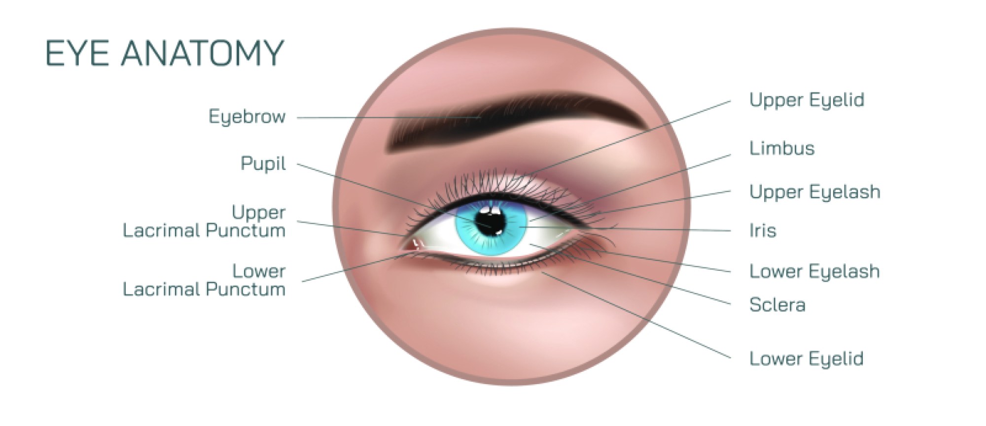
Eye Anatomy Vector Illustration Showing Human Eye Structure with Retina, Cornea, Lens, and Optic Nerve
The human eye is a highly specialized organ designed to capture light and convert it into electrical signals that the brain interprets as vision. Its intricate structure allows precise focusing, adaptation to varying light levels, and accurate perception of color, depth, and motion. A vector illustration of the human eye typically highlights key anatomical components, including the cornea, lens, retina, and optic nerve, providing a comprehensive overview of both structural organization and functional relationships. By visually mapping these elements, the diagram allows learners and healthcare professionals to understand the path of light through the eye, the mechanisms of focusing, and the transmission of visual information to the brain.
The cornea is the transparent, curved front layer of the eye that acts as the primary refractive surface. It allows light to enter the eye while simultaneously bending or refracting light rays to help focus images on the retina. In vector illustrations, the cornea is often shown as a convex, dome-shaped structure with annotations describing its curvature, thickness, and role in refractive power. Beneath the cornea lies the anterior chamber, filled with aqueous humor, which helps maintain intraocular pressure and provides nutrients to the cornea and lens. Arrows in the illustration may indicate the direction of incoming light, showing how the cornea initiates the process of focusing light toward the posterior structures. The cornea’s transparency and curvature are critical for accurate vision; any deformation or opacity can significantly impact image clarity, as seen in conditions like keratoconus or corneal scarring.
Immediately behind the cornea is the lens, a flexible, biconvex structure that fine-tunes focus for near and distant objects through a process called accommodation. Vector diagrams often depict the lens with varying curvature and position, illustrating its ability to change shape via the action of the ciliary muscles. When viewing close objects, the lens becomes more convex to increase refractive power, while for distant objects, it flattens. The lens works in conjunction with the cornea to ensure that light converges accurately on the retina, forming a sharp image. Additional annotations may highlight the suspensory ligaments (zonules of Zinn) that connect the lens to the ciliary body, emphasizing the biomechanical mechanism behind accommodation. Lens clarity is essential for vision, and conditions such as cataracts—characterized by lens opacity—can be visually represented in illustrations to show impaired light transmission.
The retina is the light-sensitive layer lining the interior of the eye, containing specialized cells that convert light into neural signals. In vector illustrations, the retina is often shown at the back of the eye, connected to the optic nerve. Two main types of photoreceptor cells are highlighted: rods, which detect low light levels and motion, and cones, which detect color and fine detail. The central portion of the retina, known as the macula, and its central depression, the fovea, are critical for sharp, detailed central vision. Arrows may indicate the pathway of light from the cornea and lens onto the photoreceptors, emphasizing the retina’s role in capturing the image and initiating the conversion into electrical signals. The illustration may also include other retinal layers, such as the bipolar and ganglion cell layers, showing the synaptic connections that transmit visual information deeper into the visual pathway.
The optic nerve transmits the electrical signals generated by the retina to the brain for processing. In vector diagrams, the optic nerve is represented as a thick cord emerging from the posterior globe of the eye, connecting to the brain’s visual cortex. The illustration may include a simplified depiction of signal transmission, showing how photoreceptor-generated action potentials travel through the ganglion cell axons to the optic chiasm, where some fibers cross to the opposite hemisphere. This pathway explains how binocular vision and depth perception are achieved. The optic nerve is also a critical clinical focus, as damage or compression, such as in glaucoma, can lead to vision loss. The vector diagram often labels the optic disc, where the nerve exits the eye, commonly referred to as the blind spot, reinforcing the anatomical relationship between structure and function.
Other supportive structures may be included in the illustration to provide a comprehensive view of eye anatomy. These include the sclera, the opaque outer coat that provides structural support; the iris, which regulates the amount of light entering the eye by adjusting the pupil diameter; the ciliary body, which controls lens shape and produces aqueous humor; and the vitreous humor, the transparent gel filling the posterior chamber that maintains the eye’s spherical shape. Vector arrows may demonstrate the flow of aqueous humor from the ciliary body through the anterior chamber and its drainage through the trabecular meshwork, providing context for intraocular pressure regulation. These additional elements underscore how anatomical components function together to maintain visual clarity, eye health, and structural integrity.
A detailed vector illustration of the human eye also often uses cutaway views to show the internal arrangement of the retina, lens, and vitreous chamber, providing a three-dimensional perspective on how light travels through the eye. The illustration may include magnified insets of photoreceptors, corneal layers, or lens fibers to highlight microscopic anatomical details. Color coding, labeling, and directional arrows make it easier for viewers to trace the path of light, understand the role of each component, and recognize potential sites of clinical concern.
By combining structural accuracy, functional pathways, and clinical context, the vector illustration of the human eye demonstrates how the cornea, lens, retina, and optic nerve work synergistically to capture, focus, and transmit visual information. The cornea and lens focus light precisely on the retina; the retina converts light into electrical signals; and the optic nerve transmits those signals to the brain for interpretation. This integrated representation helps learners appreciate the complexity of vision, the interdependence of anatomical structures, and the potential consequences of structural or functional abnormalities.
Ultimately, a vector illustration of eye anatomy provides a comprehensive educational tool that bridges the microscopic and macroscopic aspects of vision. It enables a clear understanding of how light is received, focused, and converted into neural signals, showing how each anatomical component contributes to the intricate process of sight. By visualizing the eye in this way, students, educators, and healthcare professionals can explore both normal function and pathological conditions, making the anatomy of the human eye intuitive, accessible, and clinically relevant.