Nail Layers and Structure Showing Nail Plate, Matrix, Cuticle, and Bed Anatomy in the Human Body
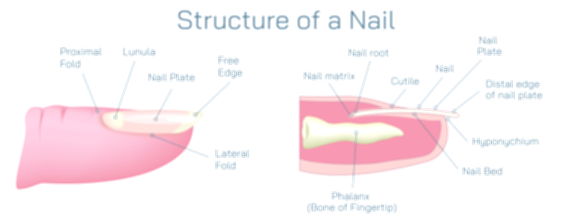
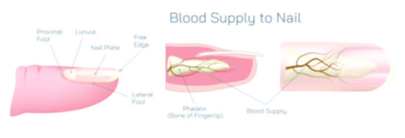
The nail is far more than a flat surface on the tip of the digit — it is a biologically engineered structure made of multiple interconnected layers that grow, protect, and reinforce the fingertips. A vector illustration showing the layers and components of the nail typically highlights the nail plate, matrix, cuticle, and nail bed, revealing how each region plays a specific role in nail growth and durability. Although much of the nail is visible externally, its true complexity lies beneath the skin surface, where specialized tissues continuously form, anchor, and guide the nail as it moves forward throughout life.
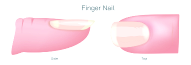
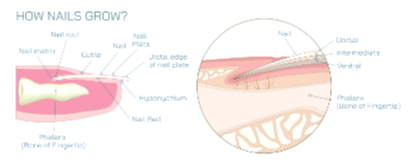
The nail plate is the hard, protective portion most familiar to the eye — the smooth, curved surface that extends over the fingertip. In an illustration, it usually appears as a multi-layered sheet composed of densely packed keratin. Rather than being a single slab of tissue, the nail plate is made of several thin layers of keratinized cells flattened and compressed together. These stacked layers give the nail strength to resist everyday forces and flexibility to prevent cracking. The nail plate is naturally translucent, which is why healthy nails appear pink — the color of the vascular nail bed beneath it. As the plate grows forward from its root at the base, older layers are constantly pushed toward the free edge, creating steady but gradual nail lengthening.
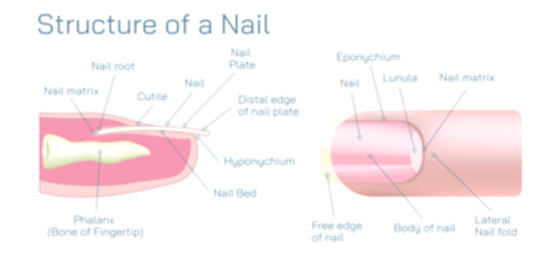
Beneath the surface lies the nail matrix, the hidden growth engine responsible for producing the nail plate. Located underneath the proximal nail fold at the base of the nail, the matrix generates new keratin cells through rapid cell division. As these newly formed cells harden and compact, they become part of the advancing nail plate. The visible white crescent at the base of some nails, called the lunula, represents the front portion of the matrix, though most of the matrix remains concealed under the skin. The shape and health of the matrix determine nail thickness, texture, and growth rate, which is why injury or inflammation in this region can lead to permanent nail deformity or growth interruption.
The cuticle, also known as the eponychium, forms a small but essential seal where the skin meets the emerging nail plate. In illustrations, it appears as a narrow band of tissue that sits at the base of the nail plate, adhering tightly to both the nail and the surrounding skin. Although often overlooked cosmetically, the cuticle plays a vital protective role: it prevents bacteria, fungi, and irritants from entering the gap between the nail plate and the underlying matrix. This biological gasket ensures the sterile environment needed for healthy nail formation. When the cuticle is damaged through over-trimming or picking, infection and inflammation can reach the sensitive matrix area, impairing nail growth.
Directly below the nail plate stretches the nail bed, the supportive and nourishing surface upon which the nail rests. The nail bed is strongly attached to the underside of the plate, keeping the nail firmly anchored while allowing uniform growth across its length. Blood vessels within the nail bed supply oxygen and nutrients to the surrounding tissues and contribute to the pinkish hue seen through a healthy translucent plate. Although the nail bed does not produce keratin, its structural alignment helps the advancing nail remain smooth and firmly positioned until it reaches the fingertip’s free edge. If the nail bed is injured or detached, the nail becomes unstable and may separate — a condition known as onycholysis.
Together, the nail plate, matrix, cuticle, and nail bed create a unified anatomical system that protects the fingertips while enabling precise movement and sensation. The nail plate acts as a shield and tool; the matrix drives growth; the cuticle guards the growth center from contamination; and the nail bed locks the plate into place and supports its movement. These layered relationships are essential for everyday functions such as grasping small objects, scratching, peeling, and enhancing tactile perception by stabilizing the skin beneath the fingertip.
A complete vector illustration typically includes:
• A side-view or cross-section cutaway showing external and internal nail layers
• The nail plate as the multilayered keratin shield on top
• The matrix positioned under the skin at the nail base, producing new nail cells
• The cuticle sealing the opening between the skin and nail surface
• The nail bed lying beneath and attached tightly to the nail plate
• Optional labeling of the lunula, nail root, and surrounding nail folds for context
Even without scientific terminology, such visuals communicate how the nail’s visible surface depends on the hidden activity of deeper structures. The nail is not merely an external covering — it is a living biological system designed for protection, movement support, and sensory enhancement. By illustrating each layer and functional component, the diagram reveals the careful coordination that allows nails to grow continuously, resist damage, and contribute to the human body’s fine motor capabilities every day.