Structure of a Nail Showing Nail Plate, Cuticle, Matrix, and Bed Anatomy in Human Biology
The human nail is often treated as a simple protective covering on the fingers and toes, yet its structure represents a seamless collaboration between several specialized tissues that allow the nail to grow, remain firmly anchored, and protect the sensitive tips of the digits while supporting fine motor function. A vector illustration of nail anatomy typically highlights the major components of the nail unit — the nail plate, cuticle, nail matrix, and nail bed — each shown as a distinct region that contributes to nail formation, strength, and adhesion to the fingertip. Although the nail appears visually flat and inert on the surface, it is a biologically dynamic system below, where active cell division and layered keratinization fuel continuous growth. Through this interplay of visible and invisible structures, the nail becomes both a protective shield and a tool enhancing grip, sensation, and dexterity.
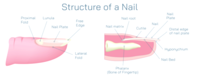
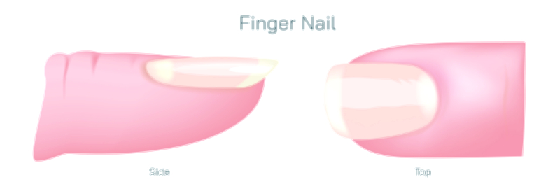
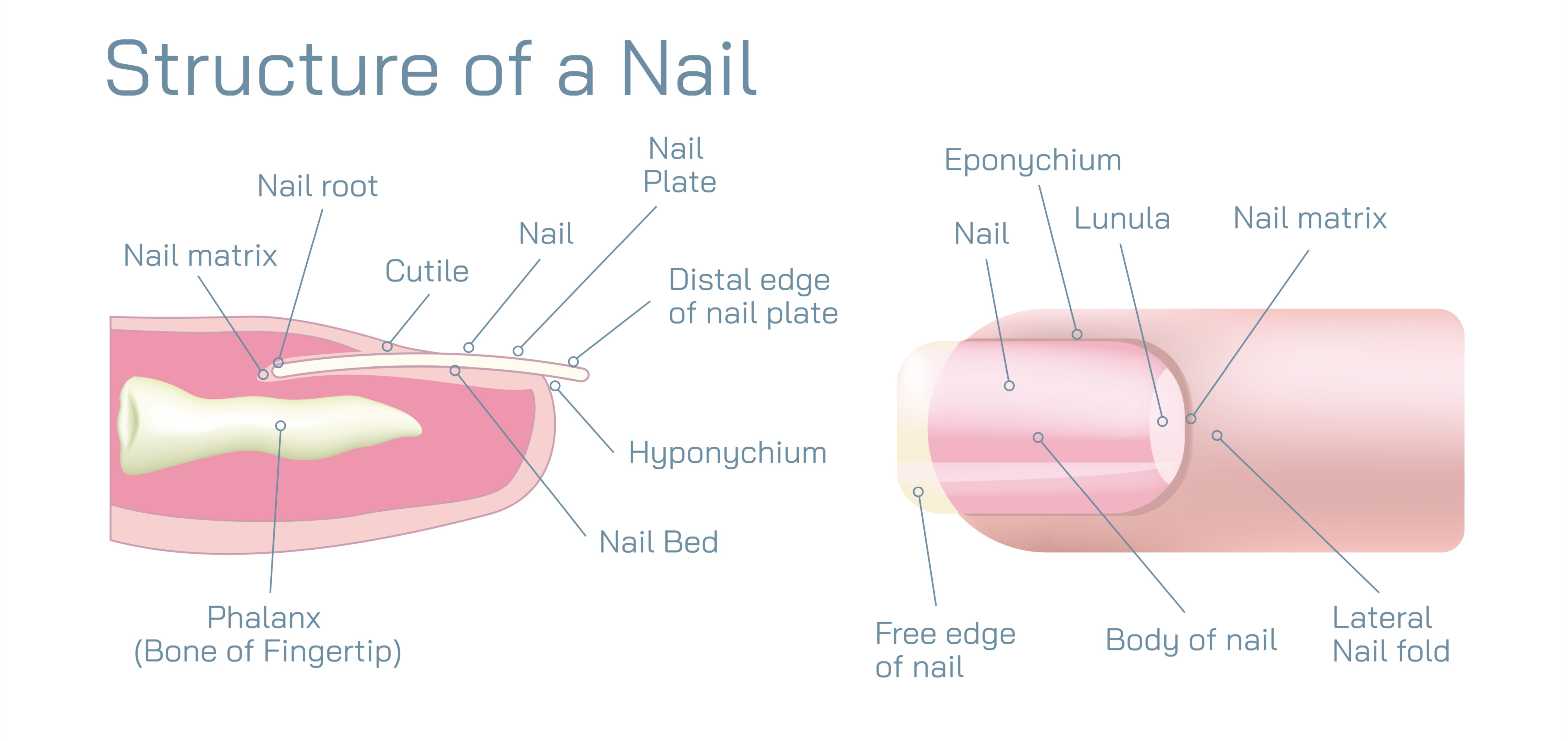
The most recognizable part of the nail is the nail plate, the hard, translucent surface that extends from the base of the finger to the free edge. In a vector illustration, the nail plate sits as the top layer of the nail unit, arching slightly to match the natural curvature of the fingertip. It is composed of keratin — a dense, fibrous protein also found in hair and the outer layer of skin — but the keratin molecules in the nail are arranged in tightly packed, flattened cells that provide hardness and durability. Because the nail plate is semi-transparent, it appears pink in healthy individuals due to blood vessels in the nail bed beneath it. The plate is made of multiple compact layers rather than a single slab of keratin, a design that gives strength while allowing modest flexibility. These layered sheets help the nail absorb small impacts without fracturing and protect the fingertip from mechanical stress, environmental exposure, and microbial intrusion.
The cuticle, sometimes called the eponychium, forms a thin but vital seal at the base of the nail plate. In an illustration, it is shown as a narrow band of skin where the nail meets the proximal nail fold. Although small in size, the cuticle creates a barrier that prevents water, debris, and infectious microorganisms from entering the space between the nail plate and the living tissue beneath. Without this natural seal, pathogens could easily reach the nail matrix and surrounding structures, leading to pain, swelling, and infection. The cuticle also helps guide the newly formed nail as it emerges from below the skin, ensuring that the plate grows smoothly and adheres properly to the nail bed. Because the cuticle acts as a biological gasket, cutting or aggressively removing it weakens this protective function, which explains why improper grooming can lead to inflammation and damage.
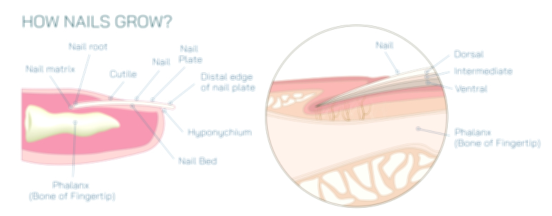
Below the surface and invisible in everyday life lies the nail matrix, the factory that produces the nail plate. It is located beneath the proximal nail fold and extends beneath the base of the nail plate as a soft, pale region shown in many illustrations. Cells in the matrix divide rapidly and gradually harden through keratinization — a process that flattens and binds them to form the tough nail. As new cells are pushed forward, the older cells compact tightly, lengthening the plate gradually from its root rather than from the tip. The visible, crescent-shaped lunula, the whitish arc near the base of the nail, marks the lower portion of the matrix in some individuals, but most of the matrix remains hidden under the skin. The matrix determines nail thickness, curvature, and growth rate, meaning injury to the matrix can permanently affect nail shape or halt nail growth. Because cell turnover here is constant, the nail grows continuously without shedding, unlike hair or outer skin layers, which go through cycles of rest and renewal.
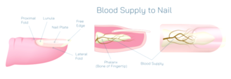
The nail bed lies directly beneath the nail plate and plays a dual role as both an anchor and a nutrient supply interface. In diagram form, it appears as the elongated, pink-tinted surface supporting the nail plate from below. The bed consists of specialized epithelium fused tightly to the plate, preventing separation and allowing the nail to feel like an integrated part of the fingertip rather than a loose surface. Capillaries in the nail bed form the color seen through the transparent nail plate and provide nourishment to the deeper nail structures. Even though the nail bed does not produce the keratin of the nail plate, it contributes to nail smoothness by keeping the underside of the plate firmly aligned across its length. Damage or disease affecting the nail bed can cause detachment (onycholysis), ridges, or discoloration, highlighting the sensitivity of the structure despite its hidden position.
Together, these components create a functional unit that adapts to the body’s mechanics. The nail protects the fingertip from pressure, abrasion, and impact, serving as a rigid backing that increases tactile sensitivity — allowing sensory receptors beneath the nail to respond more effectively when the pad presses against objects. The nail plate also supports precision tasks such as grasping tiny objects, scratching, peeling, pinching, and manipulating tools. The bed and matrix ensure that this protective tool remains anchored and capable of self-repair through continuous growth. Even tiny micro-traumas that occur during daily activity are compensated for by the relentless forward movement of the nail plate as the matrix continues generating keratin.
The anatomy of the nail also mirrors the body’s condition. Circulatory, nutritional, metabolic, and systemic health can influence nail appearance. For example, pallor can signal anemia, bluish hues may indicate low oxygen levels, ridged plates may reflect past illness or stress, and discoloration may reveal infection or systemic disease. Because the nail plate grows slowly — roughly 3–4 millimeters per month in fingernails and more slowly in toenails — changes in health may appear gradually and persist for weeks. Understanding nail structure helps medical professionals interpret these visible cues accurately rather than dismissing them as cosmetic irregularities.
A vector illustration of nail anatomy typically includes labeled cross-sections or side views to show what is normally hidden beneath the skin surface. The nail plate appears as a curved, translucent layer; the cuticle as a tight seal at the base; the matrix as the growth center sitting below the proximal nail fold; and the bed as the supportive tissue running beneath the plate. Additional features may include the nail root, the hyponychium (the skin under the free edge), and the lateral nail folds along both sides. These features collectively demonstrate that although only the nail plate appears exposed, the visible nail depends entirely on complex structures beneath and around it.
The nail is therefore not simply a rigid keratin slab but a living interface between protection and growth — a thin biological shield backed by a thriving layer of cellular machinery. The nail plate guards the digit; the cuticle seals out infection; the matrix builds and pushes the plate forward; and the nail bed anchors and nourishes the system. Together, these structures turn a fingernail into a precise, durable, and sensitive component of human anatomy. By depicting all of these relationships in one visual — surface structure, hidden growth mechanisms, and cellular interaction — a vector illustration of the nail offers not just anatomy but a window into how biology builds strength, precision, repair, and protection into one of the body’s most underestimated features.