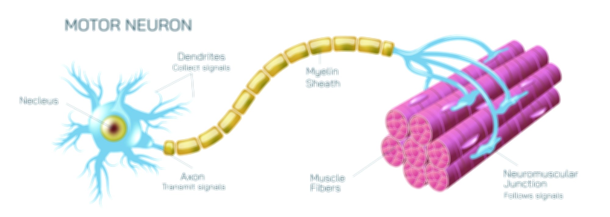
Fertilization Vector Illustration Showing Sperm-Egg Fusion, Zygote Formation, and Early Stages of Human Reproduction
Fertilization is the critical biological process that initiates human reproduction, involving the union of a sperm cell and an egg (ovum) to form a zygote, which subsequently undergoes early developmental stages leading to embryo formation. A vector illustration of fertilization typically integrates sperm motility, egg structure, fusion events, and zygote development, providing a visually intuitive and educational representation of this complex process. By combining labeled gametes, directional arrows, and developmental stages, such diagrams allow learners to understand the molecular and cellular interactions that underpin human reproduction.
At the center of the illustration is the female reproductive tract, often depicted with the fallopian tube, ovary, and uterus. A mature egg is shown released from the ovary and entering the fallopian tube, with labels identifying the zona pellucida, cytoplasm, and nucleus, which contains maternal chromosomes. Surrounding fluid in the fallopian tube may be represented with subtle shading to indicate the medium through which sperm navigate toward the egg. Arrows indicate the direction of movement of sperm from the vagina through the cervix, uterus, and into the fallopian tube, emphasizing the journey of fertilization.
The sperm cells are illustrated with distinct head, midpiece, and tail regions. Directional arrows indicate propulsion through flagellar motion, guiding the sperm toward the egg. Labels identify acrosome, which contains enzymes necessary to penetrate the egg’s protective layers, and the haploid nucleus, which carries paternal genetic material. A cluster of sperm surrounding the egg is typically depicted, showing the competition and selective nature of fertilization. Arrows or glow effects can highlight the sperm that successfully reaches and interacts with the egg.
A key component of the vector illustration is the sperm-egg fusion process. One sperm is depicted penetrating the zona pellucida of the egg, with arrows indicating enzymatic activity from the acrosome that facilitates entry. Labels such as “acrosomal reaction” and “membrane fusion” clarify the cellular mechanisms involved. Upon entry, the egg completes meiotic division, forming a female pronucleus, while the sperm forms a male pronucleus, and these structures move toward each other for fusion. Directional arrows indicate the migration and alignment of the two pronuclei within the egg cytoplasm.
The zygote formation stage is illustrated immediately following pronuclear fusion, labeled as the first diploid cell containing combined maternal and paternal chromosomes. Arrows may indicate the alignment of chromosomes on a metaphase plate prior to the first mitotic division. The illustration often shows early cell division (cleavage) as the zygote undergoes binary fission, resulting in a 2-cell, 4-cell, and 8-cell stage, eventually forming a morula. Color coding or shading may highlight individual blastomeres to show cellular progression and the uniformity of early divisions.
Vector diagrams frequently include key molecular events, such as the cortical reaction, which prevents polyspermy by altering the zona pellucida and ensuring only one sperm fertilizes the egg. Arrows and labels may indicate the release of cortical granules and the formation of a fertilization membrane, visually explaining the biological safeguards of reproduction. Additionally, illustrations may show pronuclear DNA alignment and initial mitotic spindle formation, connecting molecular processes with cellular outcomes.
The illustration may also depict fertilization in context with early embryonic transport, showing the zygote moving through the fallopian tube toward the uterus for implantation. Arrows indicate the direction of movement and the stages of cleavage along the way, linking fertilization to subsequent embryonic development. Comparative panels may show the egg before fertilization, the moment of sperm fusion, and early zygote stages, providing a temporal sequence of events.
For educational clarity, vector diagrams often use color-coded gametes and developmental stages, with labeled arrows for movement, fusion, cleavage, and pronuclear activity. Magnified insets can illustrate sperm head penetration, pronuclear formation, and early mitotic figures, providing detailed insight into cellular mechanisms while maintaining an overall structural view. Labels identify components such as egg cytoplasm, zona pellucida, sperm acrosome, pronuclei, blastomeres, and mitotic spindle, ensuring comprehensive understanding.
By combining sperm motility, egg anatomy, fusion process, zygote formation, and early cleavage stages, a fertilization vector illustration provides a complete visual understanding of human reproductive initiation. Color coding, arrows, magnified insets, and sequential panels clarify each step from gamete approach to the first divisions, connecting molecular events to macroscopic developmental outcomes.
Ultimately, a vector illustration of fertilization demonstrates the complex interplay of cellular, molecular, and mechanical processes that initiate human life. Through labeled gametes, directional arrows, pronuclear fusion, and early zygote divisions, the diagram transforms abstract reproductive biology concepts into a visually engaging, educational, and intuitive tool for medical study, biology education, and public understanding of human development.