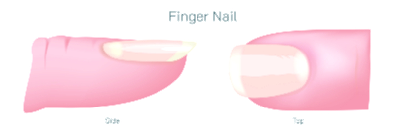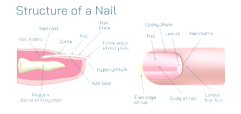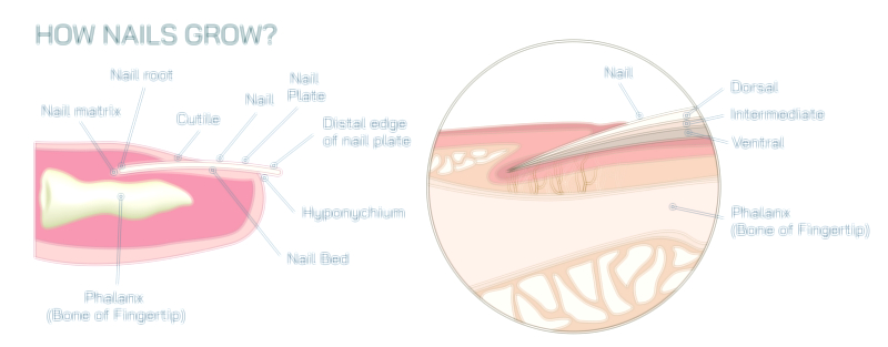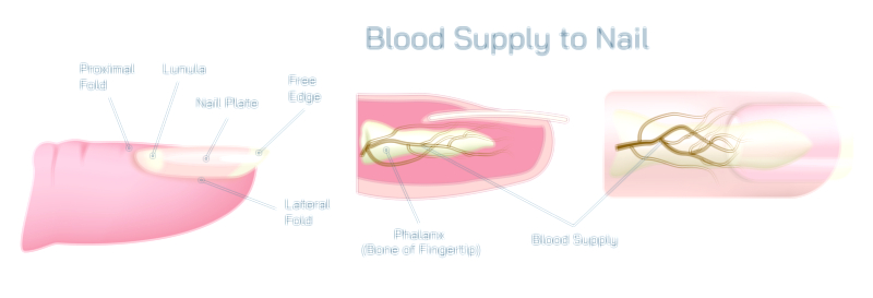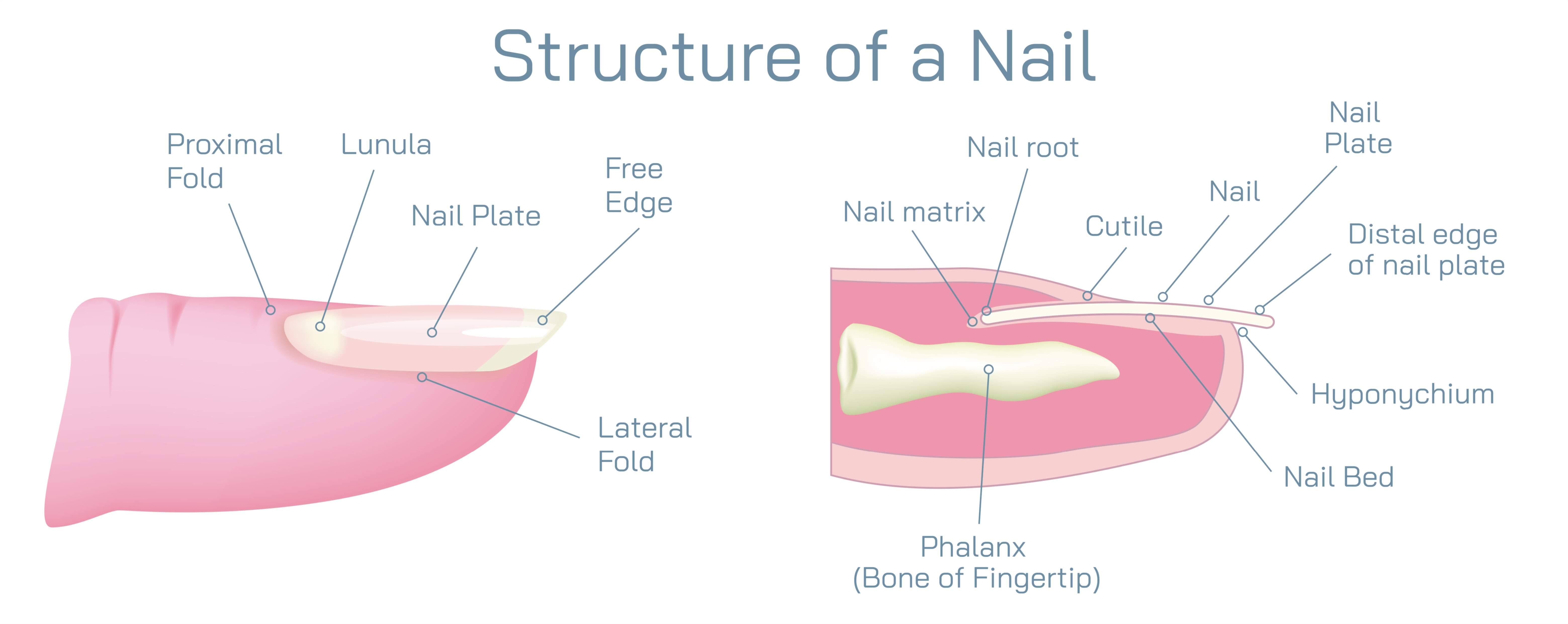
Structure of a Nail Showing Nail Plate, Cuticle, Matrix, Lunula, and Nail Bed in Human Anatomy
The human nail may look like a simple hard surface on the tip of the finger or toe, but beneath its appearance lies a remarkably coordinated biological system made of distinct anatomical components that work together to protect the digits and support fine motor function. A vector illustration of nail structure typically highlights the nail plate, cuticle, matrix, lunula, and nail bed, showing not only where each part is located but how each contributes to continuous nail growth, strength, protection, and adhesion. Visualizing these structures in cross-section allows us to see the relationship between the visible and hidden tissues that make the nail a living component rather than a passive surface.
The centerpiece of the nail unit is the nail plate, the hard, smooth, translucent structure seen externally. It is composed of densely packed layers of keratinized cells that have lost moisture and hardened into a durable sheet. Although it looks like a single piece, the nail plate is actually formed from multiple micro-layers that overlap tightly, giving the nail resilience against bending, scraping, and daily mechanical stress. Its slight curvature matches the contour of the fingertip, and because the plate is transparent, the color of the nail bed beneath gives healthy nails their pinkish tone. The nail plate is not simply a protective shield — it enables gripping, scratching, and precise manipulation of small objects because it provides solid backing to the highly sensitive fingertip skin underneath.
The cuticle, or eponychium, forms a narrow but critically important seal where the nail plate emerges from the skin at the base. In most illustrations, it appears as a thin ridge of skin attached to the surface of the nail near the proximal nail fold. Despite its small size, the cuticle serves as the gatekeeper of nail health. It closes the gap between the skin and the nail surface, preventing water, microbes, and debris from migrating toward the vulnerable tissues under the surface. When the cuticle is intact, the area beneath it remains sterile, reducing the risk of infection and inflammation. When the cuticle is damaged through over-trimming, biting, or cosmetic scraping, harmful organisms can reach deeper structures and disrupt nail growth, which is why dermatology emphasizes leaving the cuticle largely untouched.
At the foundation of the nail system lies the nail matrix, the growth center located beneath the skin at the base of the nail. It is the biological factory that produces the nail plate. Within the matrix, cells divide rapidly and undergo keratinization — the process of flattening, hardening, and losing internal fluid as they fill with tough keratin fibers. New cells formed in the matrix are pushed forward, compacting older cells tightly into the nail plate. This forward displacement explains why nails lengthen over time and why growth always moves from the base toward the fingertip rather than from the free edge backward. The appearance, thickness, curvature, and growth speed of the nail all depend on the health of the matrix. If the matrix becomes injured or scarred, the nail may grow with permanent ridges, deformities, or irregular thickness.
The lunula, sometimes visible at the base of the nail as a pale, curved crescent, represents the front portion of the nail matrix. It appears lighter because cells there are still partially keratinized and not yet fully transparent. Although the lunula is only partly visible (and in some people almost invisible), it offers a small window into the otherwise hidden matrix where nail formation begins. In a detailed illustration, the lunula is often placed between the nail plate and the deeper matrix to demonstrate its role as the transitional zone between cell creation and keratin hardening. The size of the lunula varies with finger type and individual anatomy, but its presence is one of the most recognizable indicators of the matrix’s position and activity.
Running directly beneath the nail plate is the nail bed, the tissue that anchors the plate and nourishes the surrounding structures. It contains a dense network of capillaries that provide oxygen and nutrients, and its tight bond to the underside of the plate keeps the nail smooth and firmly in place. The nail bed does not create the nail plate, but it supports and guides the plate as it advances. The pink coloration visible through the nail plate originates from the blood vessels in the nail bed. If this adhesion weakens — due to injury, infection, psoriasis, chemical irritation, or other conditions — the nail can detach from the bed, a process known as onycholysis. When the nail is properly attached, however, the nail bed acts as the stable foundation that keeps the nail plate aligned and functional.
Together, these components — plate, cuticle, matrix, lunula, and bed — form a coordinated anatomical structure designed for protection, controlled growth, and refined hand function. The nail plate shields the fingertip while enhancing touch precision. The cuticle defends the growth center from contamination. The matrix generates the keratin cells that create the nail itself. The lunula marks the most visible boundary of matrix activity and the early stage of nail formation. The nail bed holds the nail firmly in place and contributes to its coloration and stability. All of these elements function continuously and silently, ensuring that nails grow steadily at a rate of roughly three to four millimeters per month on fingers and more slowly on toes.
A complete vector illustration of nail structure typically includes:
• A side-view or cross-section diagram showing visible and hidden tissue layers
• The nail plate displayed as a multi-layered keratin sheet
• The cuticle sealing the entry point at the base of the nail plate
• The nail matrix beneath the skin, where new nail cells are generated
• The lunula as the partially visible front segment of the matrix
• The nail bed attached beneath the plate, supporting and anchoring its length
By mapping these components visually, the illustration reveals that a nail is not simply an accessory on the surface of the fingertip but a living system rooted deep beneath the skin. Its growth, protection, and mechanical function depend on the integrity of each component. When the matrix produces cells correctly, when the bed maintains adhesion, when the cuticle remains sealed, and when the nail plate stays intact, the fingertip gains a durable shield that enhances function instead of merely covering skin.
The structure of the nail embodies a design perfectly suited to human anatomy — a protective layer built from continuous growth, strengthened by keratin, stabilized by attachment to underlying tissue, and safeguarded by a natural biological barrier. This system ensures that something as seemingly simple as a nail can contribute to fine motor control, sensory improvement, and everyday protection, while reflecting internal health and adapting to the demands placed upon the hands and feet throughout life.