Central Retinal Vein Occlusion (CRVO) Eye Disease Vector Illustration Showing Retinal Blockage and Vision Loss
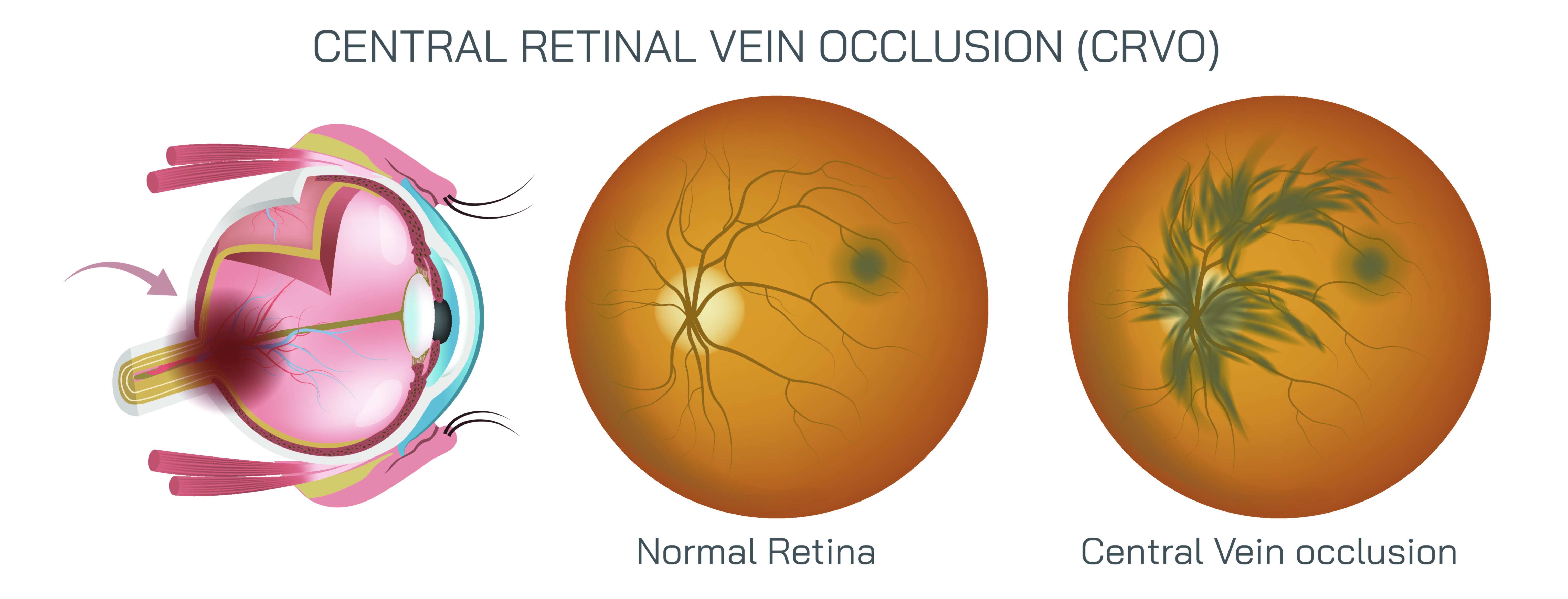
Central Retinal Vein Occlusion (CRVO) is a significant ocular vascular disorder characterized by the obstruction of the central retinal vein, leading to impaired blood drainage from the retina. This blockage causes increased venous pressure, hemorrhages, fluid leakage, and subsequent vision loss, which can range from mild to severe depending on the extent of vascular compromise. A vector illustration of CRVO typically highlights the anatomy of the retina, the central retinal vein, areas of vascular blockage, hemorrhages, and associated retinal swelling, offering a comprehensive visual understanding of the disease’s pathophysiology, clinical manifestations, and impact on vision. Such illustrations serve as crucial educational tools for ophthalmologists, optometrists, medical students, and patients seeking to understand the condition.
At the core of the illustration is the retinal anatomy, including the retina’s layers, the optic disc, and the macula. The retina is depicted as a multilayered tissue lining the posterior segment of the eye, containing photoreceptors, blood vessels, and supportive neural structures. In CRVO illustrations, the central retinal vein, which normally drains deoxygenated blood from the retina into the ophthalmic vein, is emphasized, often shown as a prominent vessel emerging from the optic disc. Arrows may indicate the direction of normal venous blood flow, while the site of occlusion is marked with a contrasting color or blockage symbol to indicate the obstruction. The illustration may also include the central retinal artery, highlighting its close anatomical proximity, as arterial compression can contribute to venous occlusion, particularly in individuals with systemic vascular risk factors.
The vascular blockage is a key feature in CRVO and is visually represented in vector diagrams by constricted or completely obstructed veins. This blockage leads to increased venous pressure, causing the thin-walled retinal capillaries to rupture, resulting in intraretinal hemorrhages. In the illustration, hemorrhages may be represented as red or dark blotches radiating from the optic disc and scattered throughout the retinal layers. Additionally, areas of retinal edema or swelling are often depicted as lightened or thickened regions, showing fluid accumulation in the macula, which is responsible for sharp central vision. This combination of hemorrhages and edema explains the visual symptoms experienced by patients, such as blurry vision, dark spots, or sudden loss of vision in one eye.
Vector illustrations also emphasize the impact of CRVO on vision by showing the affected retina in relation to the macula and optic nerve. The macula may be highlighted in a different color to indicate its susceptibility to edema, which directly affects central visual acuity. In some diagrams, vision loss is represented by shaded or blurred regions over the macula to simulate the patient’s perception, providing an intuitive connection between anatomical damage and functional impairment. The optic disc may also be labeled, as congestion at the disc is a hallmark of CRVO and helps differentiate it from other retinal vascular disorders, such as branch retinal vein occlusion (BRVO).
Causes and risk factors can also be integrated into the illustration to provide context. CRVO is often associated with systemic conditions such as hypertension, diabetes mellitus, hyperlipidemia, glaucoma, and blood clotting disorders. Vector diagrams may include simplified icons or arrows linking these risk factors to the central retinal vein, emphasizing the relationship between systemic vascular health and ocular circulation. By illustrating both local retinal changes and systemic contributors, the diagram provides a holistic view of disease mechanisms.
The types of CRVO—ischemic and non-ischemic—can be visually distinguished in vector illustrations. In ischemic CRVO, the blockage is severe, leading to extensive capillary non-perfusion, more widespread hemorrhages, and higher risk of neovascularization. Non-ischemic CRVO is milder, with partial perfusion maintained and less severe hemorrhages. Illustrations may use color intensity or shading differences to indicate the degree of perfusion and tissue damage, helping viewers understand the spectrum of disease severity and its prognostic implications.
In addition, the illustration may depict secondary complications of CRVO, including neovascular glaucoma, where abnormal blood vessel growth increases intraocular pressure, and vitreous hemorrhage, which can further compromise vision. Arrows or symbols may show abnormal vessel formation extending into the anterior segment or vitreous, connecting the pathophysiology to clinical outcomes. This aspect of the illustration emphasizes the importance of early detection and management to prevent irreversible vision loss.
Treatment-related elements may also be represented to enhance the educational value. Intravitreal injections of anti-VEGF agents, corticosteroids, or laser therapy can be indicated in vector diagrams by arrows showing the administration route or targeted area of action within the retina. These treatments aim to reduce macular edema, prevent neovascularization, and improve visual outcomes. Including treatment visualization helps contextualize the anatomical and physiological illustration in a clinical framework.
By combining retinal anatomy, vascular obstruction, hemorrhages, macular edema, optic nerve involvement, and systemic factors, a vector illustration of CRVO provides a comprehensive understanding of the disease. It highlights the dynamic interactions between blood flow, retinal structure, and visual function while emphasizing the clinical significance of timely diagnosis and management. The visual representation of vision loss alongside anatomical changes bridges the gap between pathology and patient experience, making the diagram a powerful tool for education, patient counseling, and professional reference.
Ultimately, the vector illustration demonstrates how CRVO affects the retina at multiple levels, from the microvascular changes within the central retinal vein to the macular dysfunction that impairs central vision. It integrates systemic and local factors, differentiates between types of occlusion, and conveys potential complications and treatment pathways. By presenting these elements visually and coherently, the diagram allows learners and clinicians to grasp the pathophysiology, clinical manifestations, and functional consequences of CRVO in a single, educationally effective image.