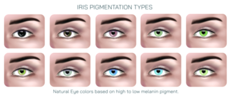
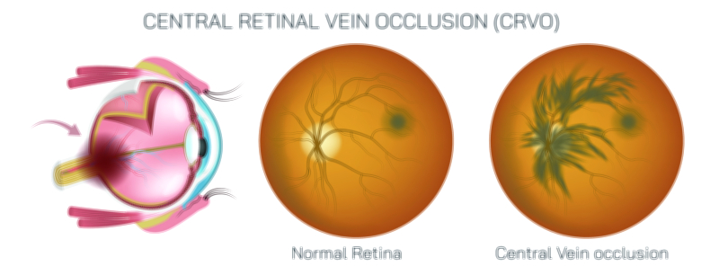
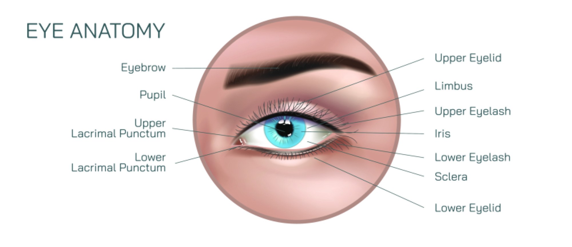
Glaucoma Eye Disease Vector Illustration for Medical and Ophthalmology Education
Glaucoma is one of the leading causes of irreversible blindness worldwide, yet it progresses so silently that many people do not realize they are losing vision until the damage is advanced. Because the disease primarily targets the optic nerve rather than the surface of the eye, it can be difficult for patients—and even students new to ophthalmology—to visualize what is happening inside the eye. A glaucoma eye disease vector illustration provides a precise and accessible way to represent the major pathological mechanisms of the condition, transforming internal and microscopic changes into understandable visual information. It becomes an extremely valuable educational asset for medical students, optometry trainees, ophthalmologists, and patient education settings because it explains not only the structure involved, but how raised intraocular pressure and other mechanisms gradually impair the optic nerve and reduce the visual field.
To understand glaucoma visually, the illustration typically begins with a clear depiction of aqueous humor flow—the fluid continuously produced inside the eye. Under normal circumstances, aqueous humor is produced by the ciliary body, circulates through the posterior and anterior chambers, exits through the trabecular meshwork, and drains through Schlemm’s canal. When this outflow pathway functions properly, intraocular pressure remains stable, supporting the structure of the eyeball without harming the optic nerve. In glaucoma, however, resistance to fluid drainage increases or the outflow system becomes blocked, causing intraocular pressure to rise. A vector diagram can show this by highlighting fluid accumulation at the level of the anterior chamber, thickened or obstructed trabecular meshwork, or narrowing of the drainage angle. Arrows showing fluid movement and obstruction deepen comprehension, especially for learners visualizing this mechanism for the first time.
Because glaucoma exists in multiple forms, educational illustrations often differentiate the two most clinically important types: open-angle glaucoma and angle-closure glaucoma. In primary open-angle glaucoma—the most common form worldwide—the drainage angle remains anatomically open, but fluid outflow through the trabecular meshwork becomes inefficient. A vector graphic can show an apparently open angle paired with thickened meshwork tissues or resistance points, helping trainees understand that the disease is not due to a physical angle blockage but to impaired outflow at the microscopic level. In contrast, angle-closure glaucoma involves an anatomically narrow or closed drainage angle, preventing aqueous humor from reaching the trabecular meshwork. A vector cross-section can help viewers see how the iris bows forward, obstructing access to the drainage system. This stark distinction is easier to grasp visually than verbally, and is essential for guiding appropriate emergency management, particularly in acute angle-closure where rapid medical intervention is critical to prevent permanent vision loss.
A well-designed glaucoma vector illustration also conveys the long-term consequences of elevated intraocular pressure on the optic nerve. As pressure increases or as optic nerve susceptibility rises (even with normal pressure in some patients), the nerve fibers within the optic disc gradually degenerate. This results in the characteristic cupping appearance seen during fundus examination—an enlarged optic cup relative to the total disc size. A vector illustration of the optic nerve head can outline the difference between a healthy nerve and one undergoing glaucomatous cupping, highlighting the thinning of the neuroretinal rim and deepening of the central cup. Such diagrams support ophthalmology students in developing pattern recognition skills for optic nerve evaluation, which is crucial because early changes can be subtle.
Another major strength of glaucoma vector illustrations is the ability to portray visual field loss. Patients do not typically experience early vision loss as blur but rather as patchy peripheral defects that gradually constrict inward. Many individuals remain unaware of this peripheral loss because the brain adapts, filling in missing areas until central vision eventually becomes affected. Illustrations that simulate the patient’s visual experience—for example, peripheral shadows, “tunnel vision,” or patchy blacked-out zones—can demonstrate how the disease impacts daily functioning while remaining deceptively unnoticed. These experiential depictions help medical professionals empathize with patients and stress the importance of routine screenings even when vision seems normal.
Educational graphics can further show associated risk factors, not as a list of text but as anatomical and physiological relationships. Factors such as aging, family history, African and Asian ancestry, myopia, diabetes, long-term corticosteroid use, and elevated intraocular pressure can be represented through complementary icons or overlays around the primary anatomy. This approach reinforces that glaucoma is both preventable through screening and manageable through timely treatment, but dangerous when ignored.
Clinical management can also be visually communicated in vector form. Treatments aim either to reduce fluid production or to enhance aqueous humor outflow. A diagram may show how medications such as prostaglandin analogs increase uveoscleral outflow, how beta-blockers and carbonic anhydrase inhibitors decrease aqueous production, or how laser trabeculoplasty improves trabecular meshwork drainage. For surgical options, vector illustrations can depict trabeculectomy creating a new drainage channel, or minimally invasive glaucoma surgery (MIGS) implants bypassing blocked pathways. Showing these treatment mechanisms within the anatomical context makes it easier for learners and patients to understand why one type of therapy is chosen over another.
Vector-format diagrams are especially valuable because of their adaptability and precision. Whether displayed in a large lecture hall, printed in ophthalmology manuals, used in clinical patient brochures, or incorporated into digital learning modules, they remain sharp and clearly readable. The ability to highlight individual structures—like the trabecular meshwork, Schlemm’s canal, ciliary processes, optic nerve head, or drainage angle—makes them suitable for layered visual learning, allowing instructors to reveal anatomical details step by step.
Glaucoma education is not only about conveying pathology but also about emphasizing urgency. Because the disease causes irreversible optic nerve damage, timely screening and adherence to treatment are crucial to preserving sight. A vector illustration communicates this urgency effectively by giving form to the damage that patients cannot physically see or feel. It reinforces that the absence of symptoms does not equal the absence of disease and that regular eye examinations—especially for high-risk individuals—are key to preventing permanent vision loss.
Ultimately, a glaucoma eye disease vector illustration functions as a comprehensive and empathetic learning tool that brings together anatomy, physiology, pathology, clinical experience, diagnosis, and treatment. It helps medical trainees build a strong understanding of the mechanisms behind the disease, aids clinicians in explaining the condition in a reassuring and effective way to patients, and supports public awareness by communicating how silent but devastating glaucoma can be when left undetected. By turning microscopic pressure changes and optic nerve degeneration into an accessible visual narrative, the illustration empowers both healthcare professionals and patients to protect vision before it is too late.