Coloboma Eye Disease Vector Illustration for Medical and Ophthalmology Education
Coloboma is a rare but clinically significant congenital eye disorder in which a portion of normal ocular tissue is missing due to incomplete closure of the embryonic optic fissure during fetal development. Because this condition can affect multiple structures of the eye—such as the iris, retina, choroid, ciliary body, macula, or optic nerve—it plays an important role in medical and ophthalmology education. A coloboma eye disease vector illustration becomes an invaluable teaching tool because it visually translates a developmental defect that originates early in gestation into clear, identifiable anatomical differences that students, clinicians, and even families can recognize. These illustrations not only clarify the physical characteristics of coloboma but also reinforce foundational concepts in embryology, ocular anatomy, and clinical ophthalmology by showing how a small disruption in fetal development can produce lasting structural and functional consequences.
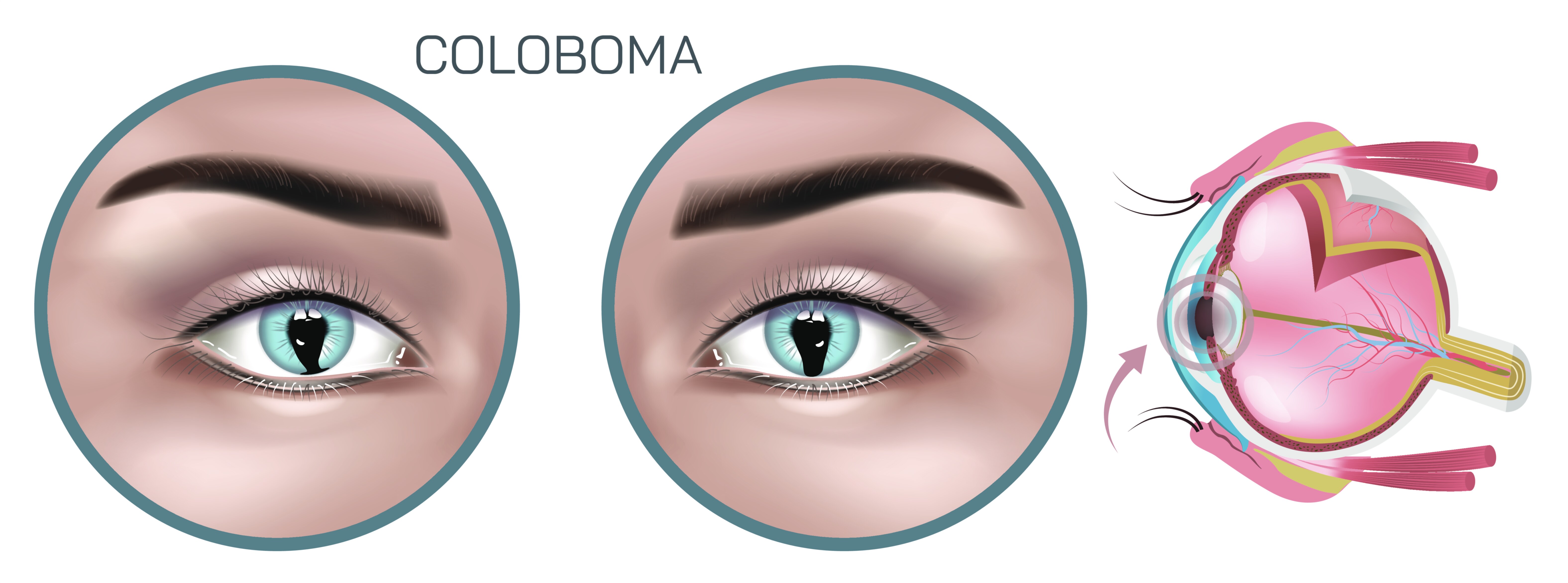
During normal eye formation, the optic fissure closes around the fifth to seventh week of gestation, sealing the developing globe. In coloboma, this closure does not complete, leaving a gap that results in missing ocular tissue. A vector illustration created for educational purposes often demonstrates this process visually by comparing normal embryonic eye development with the embryo affected by coloboma. The most recognizable presentation is iris coloboma, in which the iris has a keyhole-shaped or teardrop defect, typically oriented inferonasally. Illustrations of iris coloboma play an important pedagogical role because the abnormal pupil shape is externally visible and helps learners immediately associate the appearance with the underlying developmental mechanism. Because vector graphics provide crisp lines and smooth gradients, they allow precise depiction of the boundary between the normal iris and the missing tissue, highlighting the directional orientation that reflects the anatomical position of the original fissure.
Beyond the iris, coloboma can affect deeper ocular structures that are not visible without ophthalmologic examination. Retinal and choroidal colobomas, for example, result from incomplete closure toward the posterior segment of the fissure. These alter the architecture of the retina and can compromise the macula or optic nerve, leading to reduced visual acuity, visual field defects, and increased risk of retinal detachment. Vector illustrations depicting posterior segment colobomas are especially useful in ophthalmology education because they allow educators to visualize complex structural deficits in cross-sectional or fundus-style diagrams. The illustration might show excavation or white patches corresponding to missing retinal pigment epithelium and choroid, with clear indications of how light and neural signaling are disrupted. By reinforcing these anatomical details visually, the graphic supports a level of diagnostic understanding that is difficult to achieve through text alone.
One of the strengths of vector illustration for coloboma education is the ability to represent variation. The condition exists on a spectrum rather than as a single pattern. Some patients may only have mild iris involvement with little impact on vision, while others experience severe posterior coloboma involving the optic nerve and macula that affects visual development from early childhood. A comprehensive educational vector illustration can organize these differences into labeled panels—iris coloboma, lens coloboma, retinal/choroidal coloboma, and optic nerve coloboma—allowing medical learners to compare clinical outcomes associated with each type. This level of structured visual information allows ophthalmology residents, optometry students, and pediatric health professionals to build strong diagnostic instincts by recognizing patterns and correlating them with functional prognosis.
For learners and families, vector illustrations not only help with clinical recognition but also improve understanding of genetic and syndromic associations. Coloboma may occur as an isolated condition, but it is also seen in multisystem syndromes such as CHARGE syndrome, Trisomy 13, Cat-Eye syndrome, and others. An educational diagram that highlights this connection—perhaps through annotated side panels or iconography indicating systemic involvement—supports discussion of multidisciplinary care and genetic counseling. The precision of vector artwork helps present sensitive medical information in a format that is approachable and scientifically reliable, which is especially important when explaining lifelong conditions to families whose children have recently been diagnosed.
Vector graphics also allow powerful visual communication of functional effects. Depending on the structures involved, individuals with coloboma can experience reduced sharpness of vision, photophobia, visual field loss, depth perception difficulty, nystagmus, or significant risk of retinal detachment. A vector illustration can simulate the visual experience of a patient with coloboma—such as gaps in the visual field or impaired central focus—helping medical learners empathize with patients’ challenges rather than focusing solely on anatomical diagrams. These experiential visuals act as meaningful reminders that diagnoses carry real human consequences, influencing daily tasks, learning, safety, independence, and emotional well-being.
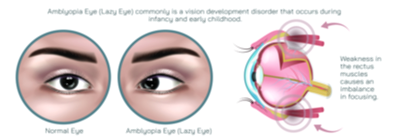
Because coloboma is congenital and non-progressive in structural origin, early identification is important for maximizing functional outcomes. Some children may benefit from visual aids such as corrective lenses, low-vision support, patching for associated amblyopia, or multilayered ophthalmologic monitoring if the posterior structures are involved. A vector illustration designed for medical education can support these discussions by depicting how therapeutic interventions aim to maximize visual potential even when structural deficits remain unchanged. The illustration becomes a storytelling tool that traces the journey from embryological origin to clinical presentation to long-term visual management.
Technically, the vector format is especially well-suited to this type of educational content due to its scalability, cleanliness, and precision. Whether printed on large classroom wall charts, projected during ophthalmology coursework, incorporated into atlas chapters, or embedded within interactive e-learning programs, the illustration remains sharp and anatomically faithful. Subtle shading can depict depth, while carefully placed highlights clarify variations in tissue absence. The graphic can further support accessibility in patient education by offering simplified versions with minimal anatomical detail for non-medical audiences and more advanced, labeled versions for clinical learners.
Ultimately, a coloboma eye disease vector illustration performs a dual role: it deepens scientific understanding and strengthens public awareness about a congenital disorder that can significantly affect lifelong vision. It bridges embryology, pediatric ophthalmology, neuro-visual development, genetics, and patient care in a single visual language that learners can reference repeatedly. By transforming a hidden developmental defect into a clear visual explanation, the illustration empowers clinicians to recognize coloboma more quickly, communicate its implications more compassionately, and participate more effectively in long-term care planning for affected individuals. Though coloboma begins as a silent interruption during fetal growth, its accurate depiction through educational vector graphics makes its mechanisms understandable, its challenges visible, and its clinical management more informed—improving the path toward better vision health for patients across their lifespan.