Cataract Eye Disease Showing Clouded Lens and Vision Impairment
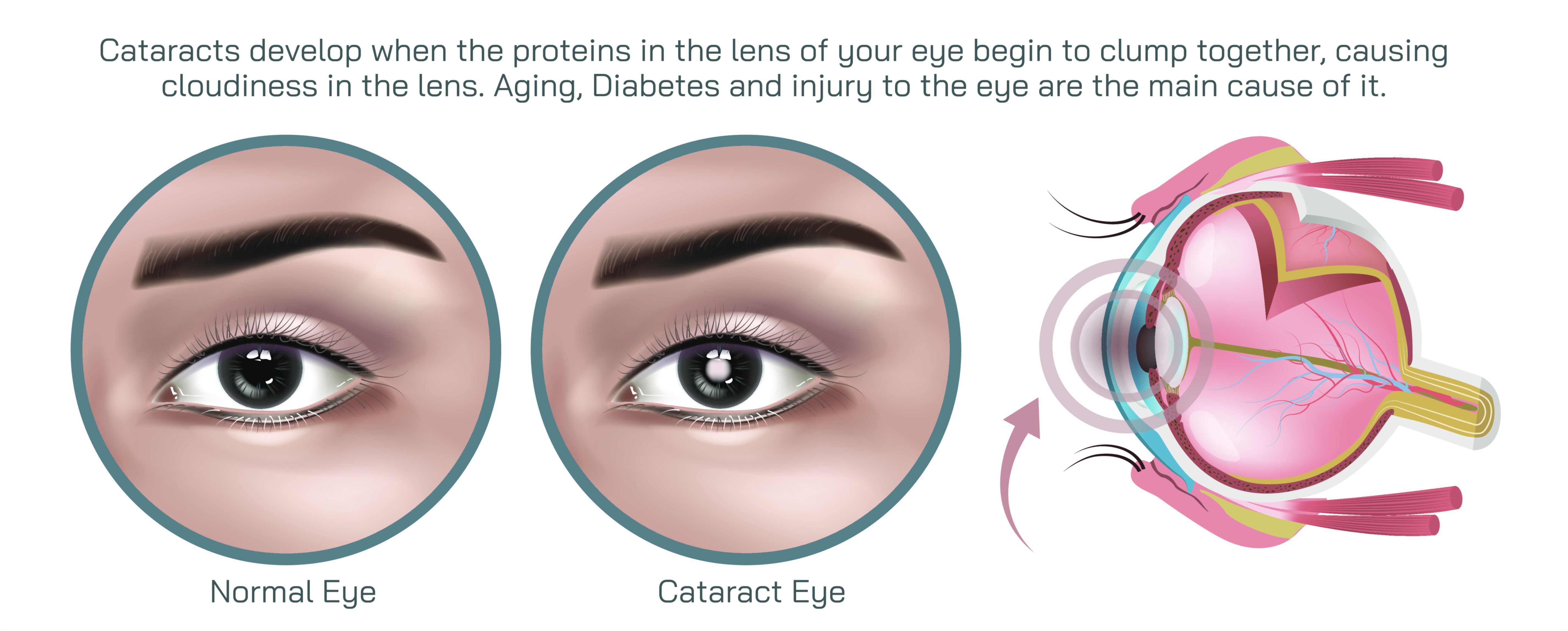
Cataract is one of the most widespread causes of vision impairment across the globe, especially in aging populations, and its defining feature is the progressive clouding of the eye’s natural lens. This clouding reduces the ability of light to pass cleanly through the lens to the retina, resulting in increasingly blurred, dimmed, or distorted vision. A medical illustration depicting cataract eye disease typically highlights an important visual contrast: a healthy eye with a crystal-clear, transparent lens versus a cataract-affected eye in which the lens appears milky, yellowish, or opacified. This visual distinction helps learners and patients immediately understand what makes cataract different from refractive errors like nearsightedness or astigmatism. The problem is not caused by the shape of the eye or the focusing muscles, but by light being obstructed before it even reaches the retina. Illustrations are therefore crucial for revealing the internal structural change that cannot be seen from outside the eye.
The healthy lens plays a vital role in clear vision: it focuses incoming light sharply onto the retina, allowing a crisp and detailed picture of the world. In cataract disease, proteins within the lens break down and begin to clump together, making the lens cloudy and less flexible. This gradual process happens over years and varies from person to person. In early stages, patients may notice only minor difficulty seeing in low light or driving at night. As the cataract worsens, the clouding intensifies, scattering and blocking light, which leads to severely blurred or hazy vision. An effective educational diagram often illustrates this light pathway: showing sharp, uninterrupted rays of light entering a healthy lens versus scattered, diffused rays struggling to pass through a clouded cataract lens. This visual pathway helps both medical students and patients understand why clarity diminishes even though the external appearance of the eye may look unchanged.
Cataracts come in different types depending on the area of the lens that becomes cloudy. A nuclear cataract, which affects the central lens, often causes gradual yellowing or browning of the vision and difficulty distinguishing colors. A cortical cataract begins at the outer edges of the lens, often showing wedge-shaped opacities that interfere with contrast sensitivity and cause glare. A posterior subcapsular cataract, located at the back of the lens, tends to produce rapid vision decline and severe light sensitivity—especially when reading or facing bright lighting. A vector illustration that maps these different opacity patterns onto the lens helps clinicians and trainees visually associate cataract subtypes with patient complaints, creating a stronger link between symptom pattern and clinical diagnosis.
Understanding vision impairment through patient-experience simulation also plays a crucial role in cataract education. People living with the condition often describe their vision as foggy, filmy, yellow-tinted, or like looking through a frosted window. They may struggle with faded colors, glare from sunlight, halos around headlights, or difficulty reading small print. Illustrations that demonstrate how cataract alters visual perception—for example, a side-by-side comparison of normal perception versus cataract-distorted scenes—help caregivers and medical educators communicate the day-to-day impact. They also help family members appreciate that a patient’s complaints are rooted in real optical limitations rather than a lack of focus, effort, or interest.
Cataract is especially important to diagnose early because patients often adapt to gradual visual loss without realizing how poorly they are seeing. For example, they may increase screen brightness, require more lighting at home, or stop driving at night without connecting these changes to declining lens clarity. Illustrations that include progressive stages—from clear lens to mildly cloudy to fully opaque—support educational discussions about when surgical referral becomes appropriate. Unlike many eye diseases, cataracts are treatable through surgery, and most individuals regain excellent vision when the clouded lens is removed and replaced with a clear artificial intraocular lens (IOL).
To support surgical understanding, vector-based graphics often show how modern cataract surgery works. Today’s treatment typically involves a procedure called phacoemulsification, in which the surgeon makes a tiny incision and uses ultrasound energy to break up the cloudy lens before removing it. An artificial intraocular lens is then implanted in its place. By portraying these steps visually—incision site, lens fragmentation, removal of lens fragments, and placement of the new IOL—illustrations help demystify surgery and reduce anxiety for patients preparing for the procedure. For ophthalmology students and residents, diagrams showing different IOL styles—monofocal, multifocal, toric, or accommodating lenses—can help reinforce how treatment can not only restore clarity but may also reduce refractive errors at the same time.
The clouding of the lens in cataract disease can be influenced by age, genetics, UV exposure, smoking, diabetes, prolonged corticosteroid use, eye trauma, or previous eye surgery. For this reason, public-facing education often includes illustrations that show both the internal changes within the lens and the lifestyle or health risk factors that contribute to cataract development. This combination deepens understanding while empowering individuals to adopt protective habits, such as UV-blocking sunglasses, smoking cessation, glycemic control for diabetes, and routine eye examinations with advancing age.
Cataract is not only a biological condition; it affects independence, mobility, communication, and emotional health. Vision loss can limit reading, driving, facial recognition, hobbies, and employment. For older adults, it can also increase fall risk and reduce social interactions. Medical illustrations that depict cataract are therefore more than diagrams—they serve as gateways to improved diagnosis, early recognition, and relief from preventable visual disability. When patients see their symptoms mirrored visually, they feel validated rather than dismissed, and when clinicians use diagrams to explain the disease, they build trust and encourage timely treatment decisions.
In essence, an educational depiction of cataract eye disease showing the clouded lens and visual impairment is a powerful instructional tool. It highlights the anatomical change at the center of the disorder, demonstrates how clouding disrupts the normal flow of light, simulates how a patient sees the world through a cataract, and connects the progression of symptoms to the decision for corrective treatment. By transforming an invisible internal problem into a clear visual story, such illustrations support medical learning, promote informed care, and help countless individuals move from visual limitation back to clarity and confidence through treatment.