Dry Eye Disease Vector Illustration for Medical and Ophthalmology Education
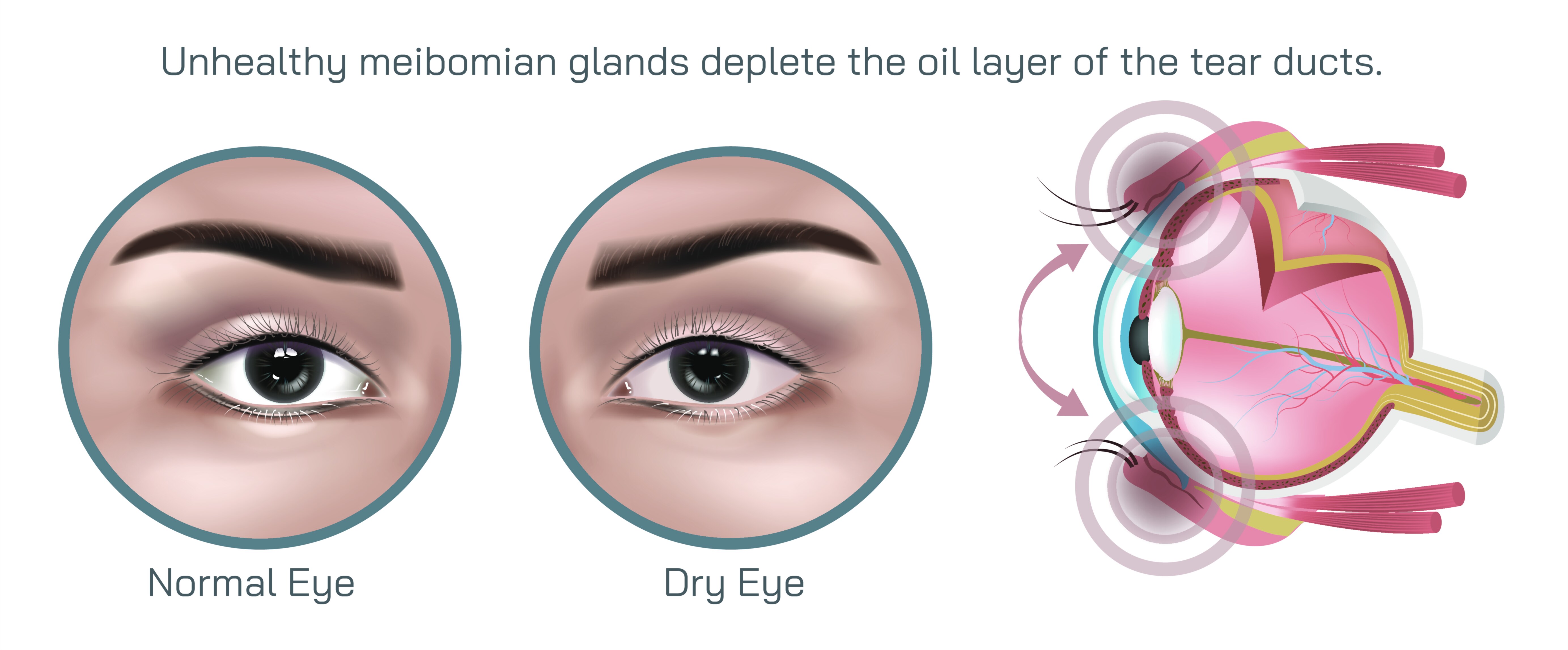
Dry eye disease is one of the most common yet often misunderstood ocular conditions, affecting millions of people across all age groups. It occurs when the surface of the eye does not receive sufficient lubrication and moisture from the tear film, resulting in discomfort, visual disturbance, and in more advanced stages, damage to the ocular surface. A vector illustration developed for medical and ophthalmology education provides a clear and structured way to explain this disorder, transforming a condition that is frequently described only through symptoms—burning, stinging, irritation, fluctuating vision—into an anatomical and physiological story that reveals precisely what is happening across the tear film, cornea, conjunctiva, and lacrimal system. Through clean lines, scalable detail, and accurate representations of the ocular surface, such an illustration becomes a critical teaching tool for doctors, medical students, eye health practitioners, and patients seeking to understand the underlying mechanisms of discomfort.
To fully convey dry eye disease visually, the illustration typically begins with an explanation of the tear film itself, which is far more complex than a simple sheet of moisture. The tear film consists of three interdependent layers: the lipid layer, produced by the meibomian glands to prevent evaporation; the aqueous layer, secreted by the lacrimal glands to supply hydration and nourishment; and the mucin layer, produced by goblet cells to ensure the tear film spreads evenly across the corneal surface. A vector illustration can depict these layers with clarity—showing how they form a cohesive barrier on the surface of the eye under normal conditions and how disruption in any one of them can trigger dry eye disease. For example, thinning of the lipid layer may lead to accelerated tear evaporation, while insufficiency of the aqueous layer results in inadequate hydration. When the mucin layer fails, the tears cannot adhere smoothly to the cornea, causing patchy dryness and fluctuating vision. These structural differences are challenging to appreciate in photographs or exams without a strong visual reference, making the vector illustration an essential teaching asset.
Because dry eye disease is multifactorial, the illustration often divides the condition into two primary categories to support clinical reasoning. Evaporative dry eye, the most common form, is primarily linked to meibomian gland dysfunction, in which the glands become blocked or their secretions deteriorate in quality, reducing the protective lipid layer. A vector graphic can highlight enlarged or obstructed meibomian glands, stagnated secretions, and a dangerously thinning lipid surface that allows rapid tear loss. The second category, aqueous-deficient dry eye, occurs when lacrimal gland output decreases—often due to aging, hormonal changes, autoimmune conditions like Sjögren’s syndrome, or long-term contact lens wear. Here, the vector illustration may emphasize reduced tear volume, dryness of the conjunctiva, and corneal exposure. When combined, these portrayals help educators map symptoms back to root causes and guide treatment pathways in both ophthalmology and optometry training.
The clinical consequences of dry eye disease can also be communicated visually in a vector diagram, showing how the condition progresses if left untreated. Persistent dryness causes micro-abrasions on the corneal epithelium, triggering inflammation that further destabilizes the tear film and perpetuates the cycle of irritation. Redness, friction from blinking, and punctate epithelial erosions are concepts far easier to learn when shown graphically rather than only described. Students and patients are able to understand why the disease produces sensations such as grittiness, burning, foreign body feeling, or difficulty maintaining comfortable visual focus for prolonged reading or computer work. The vector illustration can simulate optical disturbances—such as fluctuating clarity, glare, halos, or increased sensitivity to wind and air conditioning—to reflect how dry eye impacts daily life beyond the microscope lens.
One of the most valuable contributions of vector illustrations to dry eye education is helping viewers appreciate that this is not simply an inconvenience but a true chronic medical condition with systemic and behavioral associations. The illustration can draw attention to the relationship between prolonged digital screen use and decreased blink rate, showing how incomplete blinking fails to distribute tears evenly, quickly destabilizing the tear film. Likewise, visual cues might highlight the effects of contact lens wear, environmental stressors like low humidity or air pollution, and systemic contributors such as hormonal changes and autoimmune disorders. Through this full-spectrum approach, the illustration becomes a holistic learning instrument, helping clinicians make more informed differential diagnoses and allowing patients to recognize factors they can modify in their daily environment.
The graphic also plays a foundational role in explaining treatment and preventive strategies. Clinicians may use it to show where therapeutic interventions act within the ocular system—whether artificial tears support the aqueous layer, warm compresses assist meibomian gland secretion, omega-3 supplementation influences lipid production, punctal plugs reduce tear drainage, or anti-inflammatory medications break the cycle of surface irritation. The vector format enables the layering of educational overlays around the anatomical core, so learners can see not only “what” is being treated but also “why.” For patients who struggle to understand the need for consistent care in a chronic condition that fluctuates day to day, this visual clarity reinforces long-term treatment adherence and reduces frustration.
The versatility of a vector illustration becomes especially relevant in ophthalmology, where precise scaling is essential to convey microstructures correctly. The same graphic can be enlarged for projection in a residency training lecture or minimized for display in an optometry pamphlet without losing detail. This consistency helps standardize medical education across institutions and supports communication between specialists, general practitioners, and patients.
Ultimately, a dry eye disease vector illustration is far more than a visual representation of symptoms—it is a concentrated delivery of anatomical, physiological, and clinical understanding. It helps future ophthalmologists and optometrists build diagnostic intuition, equips medical educators with a clear teaching reference, guides patients toward more meaningful participation in their treatment, and increases awareness of a condition many underestimate until it affects quality of life. By transforming the invisible instability of the tear film into a clear and comprehensible visual narrative, the illustration strengthens the connection between scientific knowledge and clinical care, supporting earlier diagnosis, patient empowerment, and healthier lifelong vision.