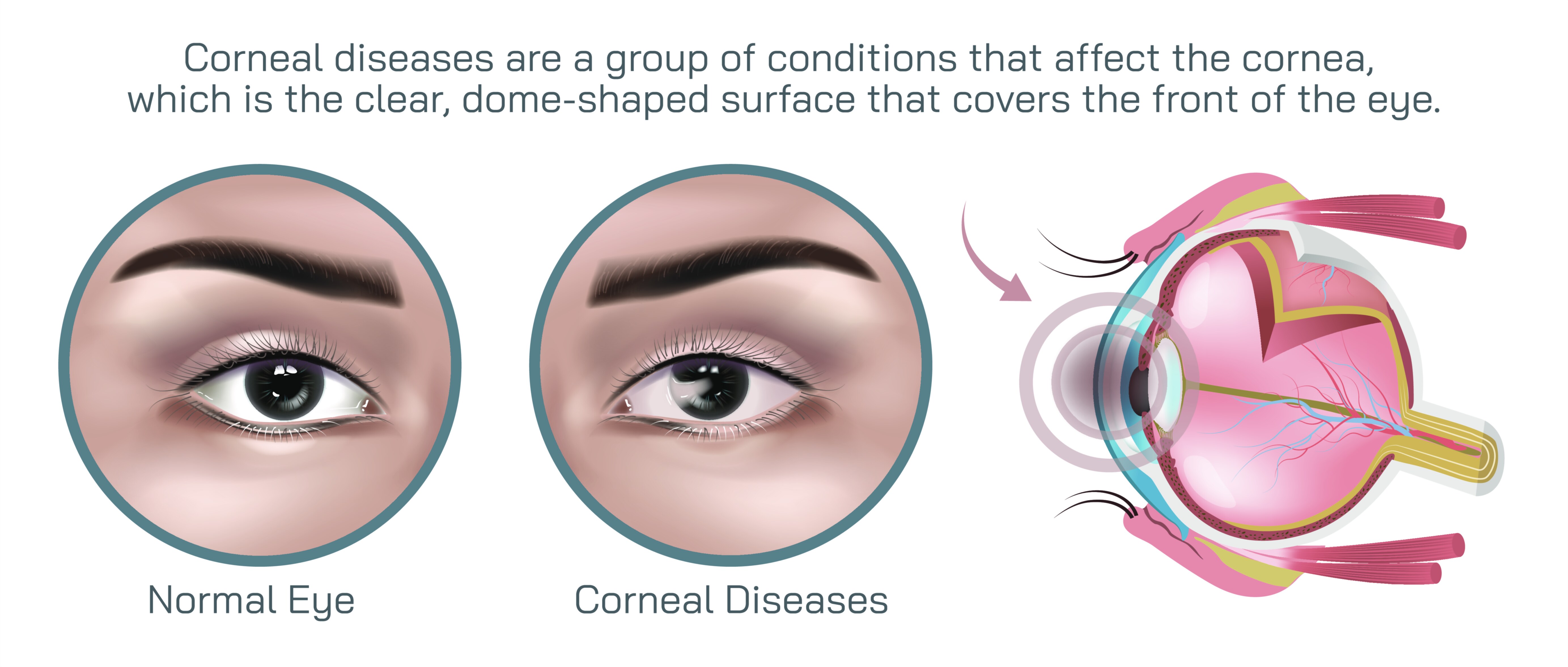
Corneal Dystrophy Eye Disease Vector Illustration for Medical and Ophthalmology Education
Corneal dystrophy is a diverse group of inherited eye disorders characterized by the gradual accumulation of abnormal deposits within the layers of the cornea, leading to progressive changes in clarity, structure, and visual performance. Because the cornea is responsible for refracting light onto the retina with precision, even minor disruption to its transparency or smoothness can result in significant visual impairment. A vector illustration designed for medical and ophthalmology education plays a particularly important role in making this disease category easier to understand, because corneal dystrophies are not only complex in their biological origins but highly variable in presentation. Such an illustration visually decodes the corneal layers—epithelium, Bowman’s layer, stroma, Descemet’s membrane, and endothelium—and maps how different dystrophies affect specific anatomical regions. This type of graphic supports both students and trained clinicians by matching theoretical knowledge with a precise visual reference, which is critical for distinguishing between similar ophthalmologic conditions that require different diagnostic and therapeutic approaches.
Modern ophthalmology recognizes that corneal dystrophy is not a single disease, but more accurately a classification of multiple disorders, each with distinct visual patterns, inheritance patterns, and structural pathology. Yet from the patient’s perspective, early symptoms often appear deceptively simple: recurrent eye discomfort, fluctuating blurred vision, foreign body sensation, and increased sensitivity to light. Over time, as abnormal materials like lipids, proteins, or calcium accumulate within the corneal layers, transparency progressively decreases. A vector illustration that highlights both normal and diseased corneal anatomy gives educators a structured visual means to demonstrate how a perfectly smooth and transparent cornea transitions into tissue marked by opacity, roughness, swelling, and surface irregularities. In medical school and residency programs, these visuals help learners anchor their understanding in anatomy rather than relying solely on verbal descriptions or case images, which can be highly variable due to lighting and imaging differences.
Educational illustrations also become invaluable when demonstrating the diversity among dystrophy subtypes. For example, epithelial basement membrane dystrophy affects the outermost layers and is associated with irregular maps, dots, and fingerprint-like epithelial patterns. Granular dystrophy presents as breadcrumb-like white opacities within the stroma, while lattice dystrophy shows amyloid deposits arranged in lattice-shaped linear branching patterns. Fuchs endothelial dystrophy affects the posterior corneal layers and is characterized by guttata—wart-like excrescences on Descemet’s membrane—which eventually lead to stromal edema due to endothelial pump failure. A vector graphic can depict these unique patterns with accuracy and consistency, allowing ophthalmology trainees to compare and memorize the diagnostic clues that differentiate them. This type of side-by-side visual classification not only reinforces disease recognition, but also indicates how lesion location influences prognosis and treatment strategy.
Another reason vector illustrations are particularly effective for teaching corneal dystrophy is their ability to demonstrate progressive change over time. Corneal dystrophies characteristically worsen slowly, often beginning in childhood or early adulthood and progressing across decades. Patients may experience symptom-free intervals followed by periods of episodic pain related to recurrent corneal erosions. These erosions are caused by unstable epithelial attachment and cause acute discomfort when the eye opens after sleep. The ability to present a sequence of disease stages within a clean vector diagram assists in teaching that corneal dystrophies should not be interpreted as static disorders. Instead, they reflect evolving pathology that moves across distinct phases—from minor deposits that cause little disturbance to severe opacities that disturb daily life and, ultimately, to the possibility of significant visual loss requiring advanced clinical intervention.
Treatment perspectives also deepen educational relevance, and vector illustration supports this by enabling clear demonstration of how medical and surgical strategies target affected corneal layers. For example, hypertonic saline, lubricating drops, therapeutic bandage contact lenses, and epithelial debridement may be recommended for early-stage epithelial dystrophies with erosions. Phototherapeutic keratectomy, which uses excimer laser smoothing of Bowman’s layer and anterior stroma, is particularly effective for certain dystrophies like granular and epithelial basement membrane dystrophy. In contrast, endothelial dystrophies such as Fuchs often require surgical techniques that selectively replace the dysfunctional endothelial layer through Descemet stripping endothelial keratoplasty or Descemet membrane endothelial keratoplasty. A vector illustration can depict these interventions visually, clarifying why treatment differs depending on whether pathology lies in the epithelium, stroma, or endothelium, and reinforcing the idea that dystrophy classification directly determines therapeutic planning.
Vector illustrations further support ophthalmology education by making implicit concepts—like optical distortion—explicit and relatable. The smooth, glass-like surface of a healthy cornea ensures predictable refraction, while diseased corneal textures scatter and block light. Graphic depictions of glare, halos around lights, shadowed regions of vision, or patchy blurring help medical trainees and caregivers understand how patients experience the disease on a daily basis. For those living with corneal dystrophy, symptoms can include difficulty driving at night, inability to read comfortably, visual fatigue due to poor contrast sensitivity, and fluctuations in clarity depending on corneal hydration. Seeing these distortions simulated in visual educational material helps caregivers and clinicians appreciate the disorder not merely as something observed through a slit-lamp exam, but as a condition affecting a person’s daily autonomy, emotional well-being, and sense of stability in their visual world.
Because corneal dystrophy is primarily genetic, patient and family counseling are essential aspects of medical care. A vector illustration assists this process by presenting anatomical changes in a manner that is easy for non-specialists to understand, reducing anxiety associated with unfamiliar terminology. Families may be unaware that early screening is beneficial for relatives who are at higher risk. Educational diagrams explaining inheritance patterns and age-related progression help patients recognize why regular follow-up visits are recommended even during symptom-free periods. The visual clarity of vector diagrams is especially helpful when discussing why seemingly sudden symptoms such as morning pain or foggy vision are not random but reflect diagnostic hallmarks that physicians monitor carefully during examinations.
In addition, because ophthalmology is increasingly globalized, the use of vector illustrations enables standardization of medical teaching across borders. Corneal dystrophy case photographs can vary drastically in color balance, lighting, and magnification, which sometimes makes anatomical teaching inconsistent across institutions. Vector graphics, in contrast, provide clean, reproducible, and universally consistent representations of pathology that support both foundational learning and continuing professional education. Whether appearing in digital textbooks, web-based lectures, posters in clinical training centers, or interactive ophthalmology simulators, the illustration retains sharpness and detail without degradation, allowing learners to focus exclusively on diagnostic structure rather than image quality limitations.
Ultimately, a corneal dystrophy eye disease vector illustration performs a role far greater than visual labeling. It brings together anatomy, pathology, genetics, clinical presentation, symptom experience, and treatment planning into a single educational resource that enables deeper understanding at multiple learning levels. For medical trainees, it reinforces the need to identify subtle slit-lamp findings and correlate them with functional symptoms. For ophthalmology residents, it supports differential diagnosis between multiple dystrophy subtypes. For surgeons, it helps conceptualize when keratoplasty becomes necessary and which layers should be targeted. For patients and caregivers, it demystifies the disease and empowers collaborative decision-making. By transforming complex microscopic changes in the corneal tissue into precise, accessible visual knowledge, the illustration helps convert scientific understanding into better diagnosis, timely intervention, improved quality of life, and ultimately stronger vision health outcomes for those affected by this lifelong group of conditions.