Amblyopia Eye Disease Vector Illustration for Medical and Vision Health Education
Amblyopia, commonly known as “lazy eye,” is one of the most significant visual development disorders in childhood and carries lifelong implications if not detected early, making it a critical topic in both medical education and vision health awareness. A vector illustration designed to portray amblyopia for educational purposes takes on an important responsibility: translating a neurological condition that is often invisible to the naked eye into a clear, structured image that communicates its mechanism, impact, and urgency of treatment. Unlike many eye diseases that involve physical tissue damage or anatomical deformation, amblyopia is rooted in the disruption of communication between the eye and the brain. It is a vision development disorder in which one eye fails to achieve normal visual acuity because the brain begins to favor the signals from the stronger eye, gradually suppressing or ignoring input from the weaker one. The affected eye may appear perfectly healthy, so without proper screening, the condition often goes unnoticed. This makes the use of visual medical illustration vital for teaching doctors, students, parents, and the general public about what is happening beneath the surface and why early diagnosis matters.
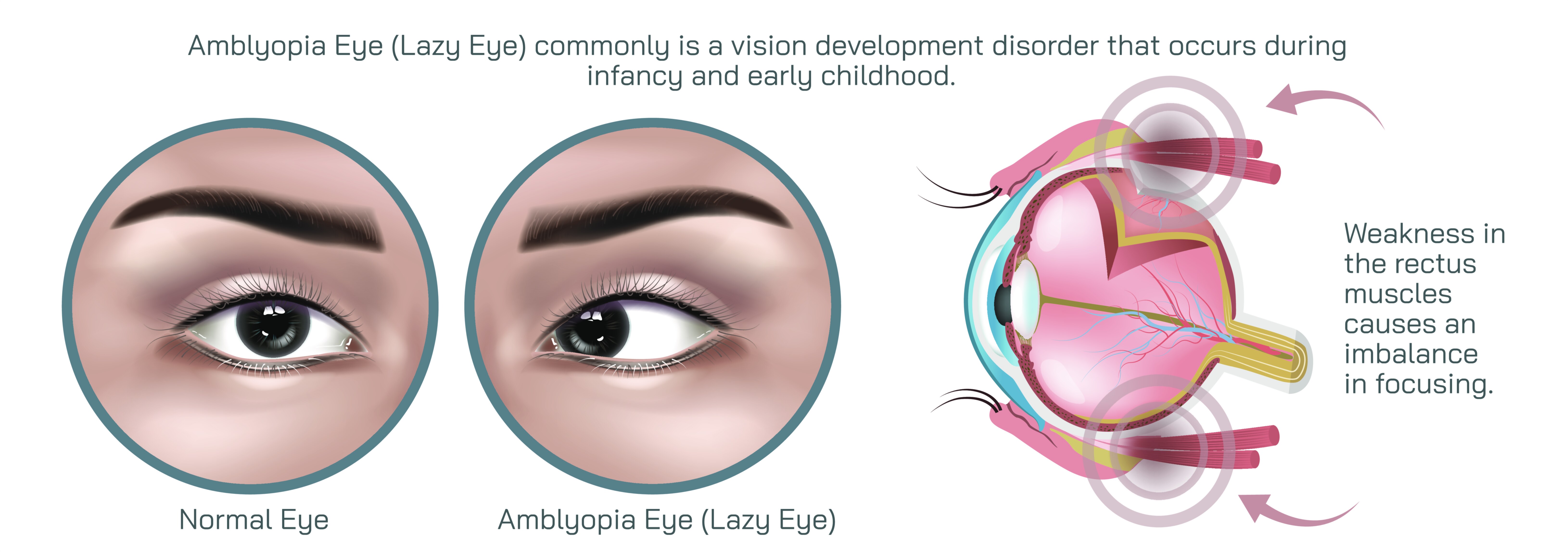
A vector illustration crafted for amblyopia education typically highlights the contrast between a normally functioning visual system and one affected by the disorder. In a typical visual pathway, each eye sends well-focused signals to the brain, allowing the brain to fuse both images into a single, sharp three-dimensional perception of the world. In amblyopia, however, the brain receives one clear image from the dominant eye and one blurred or misaligned image from the weaker eye. The brain, in its natural tendency to simplify input, begins to disregard the inferior signals from the affected eye. Over time, this causes weakening of neural pathways connected to that eye, diminishing its role in vision even though the eye itself remains structurally capable of seeing. A vector graphic that depicts weakening neural pathways, fading signal lines, or diminished detail in the perceived image provides learners with a concrete depiction of the suppression process occurring at the level of the brain rather than the surface of the eye.
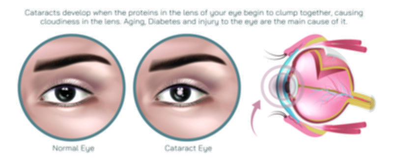
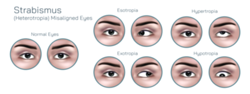
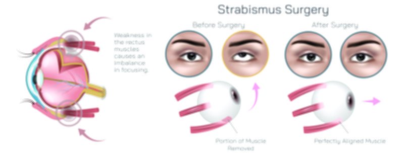
Since amblyopia emerges in different forms depending on the underlying cause, educational illustrations often differentiate between its major types. One category is strabismic amblyopia, caused by eye misalignment, where the brain struggles to merge the two different visual inputs and suppresses the deviated eye to avoid double vision. Another is refractive amblyopia, arising when there is a large difference in refractive power between the eyes, causing chronic blur in one eye that the brain learns to ignore. A third category is deprivation amblyopia, which occurs when something obstructs light from entering the developing eye, such as drooping eyelids or congenital cataracts. A vector illustration that includes these variations helps medical and health students understand that amblyopia is not a single event but a range of mechanisms leading to the same neurological outcome. This visual separation of causes allows for deeper comprehension of diagnostic pathways and treatment decisions because the type of amblyopia influences how therapy should be approached.
One of the reasons vector illustration is ideal for medical education is its ability to present specific visual cues without distortion. Demonstrating a blurred image as perceived through the amblyopic eye, reduced contrast sensitivity, difficulty with fine detail recognition, and disruption of depth perception can be visually represented in a way that photographs cannot always accomplish. Many children with amblyopia do not complain about their vision because the dominant eye compensates, and they may not realize that their perception is different from others. Seeing a vector representation of normal vision next to amblyopic vision conveys to educators and caregivers the importance of early screenings so that the condition does not silently progress. Moreover, the scalability of a vector graphic makes it possible to use it effectively in many formats—from posters in pediatric clinics and optometry offices to interactive slides in ophthalmology programs and anatomy e-learning courses. This ensures that the didactic message is communicated consistently, regardless of display size.
Educational materials that include vector illustrations are especially helpful in explaining how amblyopia affects life if left untreated. Children may struggle with depth perception, which can influence motor coordination, sports performance, and overall confidence. Fine motor tasks and reading speed may be affected, and in adulthood, the loss of binocular vision can limit career options in fields requiring precise stereoscopic vision. A high-quality vector depiction of stereopsis impairment and eye-brain disconnect helps clinicians explain these outcomes without overwhelming viewers, especially when speaking to parents who may be unfamiliar with medical terminology. When parents visually grasp what is happening inside the child’s visual system, they tend to take screening and treatment recommendations much more seriously, improving prevention outcomes in the broader population.
Amblyopia is unique among eye disorders because it is highly treatable and sometimes fully reversible if addressed early. During childhood, the brain has strong neuroplasticity, meaning neural connections can be strengthened or reorganized when the weaker eye is stimulated. A vector illustration representing therapy success—for example, increased neural activity between the previously suppressed eye and the visual cortex—can help communicate the healing process. Treatments such as corrective lenses, patching the dominant eye, atropine drops to blur the stronger eye temporarily, or modern digital visual exercises are most effective when initiated early, often before age seven. Showing this visually in educational materials reassures families that treatment is both effective and time-sensitive, motivating timely intervention and long-term commitment to therapy.
Within medical and vision science education, vector illustrations of amblyopia also serve a neuroscientific purpose beyond clinical care. They allow students to see vision not simply as a function of the eyes but as a complex sensory process dependent on normal brain development. The illustrations often highlight the route from the retina to the visual cortex, providing insight into how structural health of the eye is not always equivalent to functional vision. This recognition helps students appreciate how developmental disorders can produce significant impairment without obvious damage to observable anatomy. It also fosters interdisciplinary collaboration among ophthalmologists, neurologists, pediatricians, vision therapists, and optometrists, strengthening the understanding that healthy vision depends on both optical clarity and neural processing.
The emotional dimension of amblyopia education should not be overlooked, especially since the condition overwhelmingly affects children during key developmental stages. An illustration that communicates the disorder with calm, supportive visual language can reduce anxiety for both children and caregivers, making them more receptive to treatment. For young audiences, simplified and gentle visuals can make the topic approachable, whereas more detailed anatomical diagrams serve advanced learners in clinical environments. The versatility of vector design allows both styles to reflect the same underlying medical message while adapting to different contexts and age groups effectively.
In the realm of public health, amblyopia is one of the most preventable causes of long-term vision impairment. The power of the vector illustration lies not just in explaining how the disease develops, but in motivating action—eye screenings in early childhood, vision checks before school enrollment, awareness of risk factors in premature infants or children with a family history of strabismus, and compliance with treatment. When scientific knowledge is visually distilled into an educational format, the condition becomes less abstract and more actionable. Instead of remaining a hidden developmental disorder that goes undiagnosed until it becomes irreversible, amblyopia becomes something that caregivers recognize, child health programs prioritize, and medical institutions treat early enough to prevent lasting damage.
Thus, an amblyopia eye disease vector illustration becomes much more than an image; it is a comprehensive educational tool bridging anatomy, neurology, ophthalmology, child development, and public awareness. It helps future medical professionals understand diagnostic mechanisms, guides treatment discussions with patients and caregivers, and carries the collective mission of reducing preventable vision loss in children across the world. Through visually communicating the suppressed eye–brain connection and the hope of recovery through early intervention, the illustration encourages both understanding and empowerment—turning medical knowledge into a meaningful pathway for better lifelong vision health.