Amblyopia Eye Disease Vector Illustration for Medical and Eye Health Education
Amblyopia, widely recognized as “lazy eye,” is one of the most important visual disorders to understand in medical, pediatric, and eye health education because it affects visual development during childhood when the brain is still forming neural pathways that control sight. A vector illustration created to represent amblyopia for educational purposes plays a vital role in teaching how the disease develops, how it alters the visual system, and why early diagnosis is critical for preventing lifelong impairment. Unlike many ocular diseases that originate from structural damage in the eye, amblyopia is primarily a developmental neurological condition in which one eye fails to establish proper communication with the visual cortex. The eye itself may appear anatomically normal, yet the brain gradually suppresses visual signals from it, leading to reduced vision that cannot be corrected simply with glasses unless intervention occurs early. This complexity makes visual medical illustrations particularly valuable because they help students, parents, and healthcare providers grasp what is happening beneath the surface.
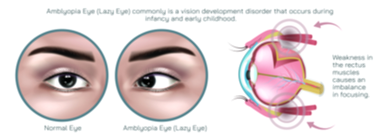
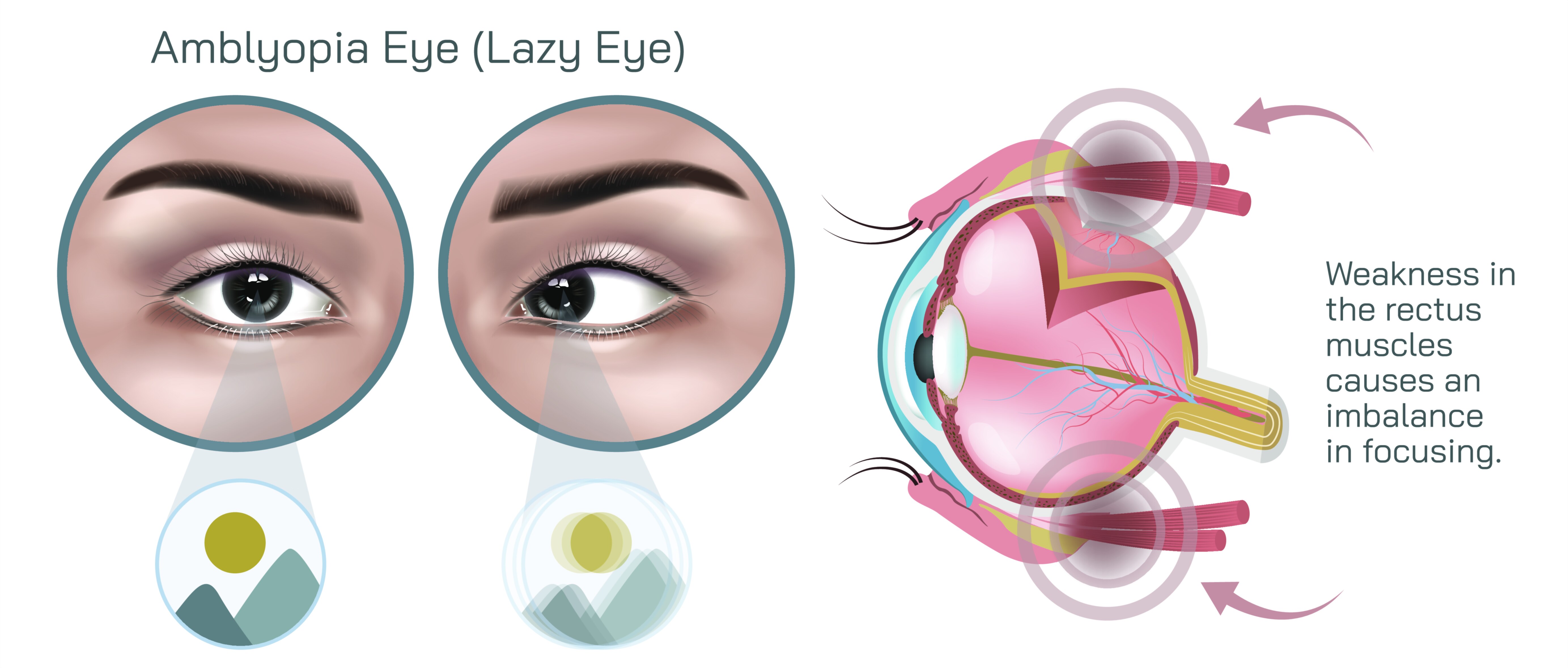
A vector illustration designed for amblyopia education typically contrasts the normal visual pathway with the affected one. In a normally developing child, both eyes send clear and balanced signals to the brain, which fuses the two images into sharp binocular vision. In amblyopia, one eye sends weaker, blurred, or misaligned signals, and the brain begins to favor the stronger eye. The weaker eye’s neural pathway becomes under-stimulated and underdeveloped, eventually causing functional visual loss even though the eye structure remains intact. A precise vector graphic can show this suppression visually with diminished signal lines from one eye, asymmetry of focus, or contrasting clarity between the two visual channels. Because amblyopia is an invisible disorder to the untrained eye—there is no obvious external sign in many cases—such illustrations transform conceptual understanding into tangible educational insight.
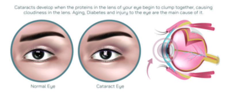
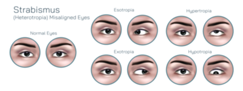
Different causes of amblyopia can be represented in educational illustrations depending on the purpose. Strabismic amblyopia arises when one eye turns inward, outward, upward, or downward, preventing the brain from aligning images from both eyes. Refractive amblyopia occurs when there is a significant difference in refractive error (such as farsightedness, nearsightedness, or astigmatism) between the two eyes, causing one eye to produce a chronically blurred image. Deprivation amblyopia results from obstruction of vision during infancy or early childhood, such as due to congenital cataracts or ptosis (drooping eyelid). A vector illustration that differentiates these patterns helps educators explain not only that amblyopia affects vision but also how different clinical triggers interfere with neural development. In medical training, this visual clarity supports quicker diagnostic thinking because pattern recognition becomes instinctive when backed by strong visual memory.
The benefit of using vector graphics for amblyopia education lies in scalability and precision. Whether displayed in a children’s eye health campaign poster, a medical school textbook, an optometrist’s office pamphlet, or an ophthalmology conference presentation, the illustration needs to remain sharp at every size. Vector design ensures that lines representing visual pathways, anatomical structures, or comparative retinal images retain crisp clarity, allowing learners to focus on key concepts without distortion. The scalable nature also enables adaptation for animated simulations, digital learning modules, and interactive screens in telemedicine platforms, where illustrating the functional difference between the amblyopic eye and the stronger eye improves patient comprehension.
For broader public awareness, especially among parents, an educational vector illustration showing how a child sees with amblyopia can carry major preventive value. When parents understand that gradual suppression of vision may not present obvious symptoms—because the child relies on the stronger eye—they become more likely to prioritize early childhood eye screening. Many amblyopia-focused illustrations therefore depict a side-by-side comparison: crisp, focused vision through a normal visual system versus blurred or suppressed vision through an amblyopic one. Feature cues such as washed-out details, low contrast, uneven focus, and depth perception issues can be shown graphically to demonstrate the real-world visual experience of a child living with untreated amblyopia. This helps convey that kids may not complain because they do not realize something is wrong—they simply adapt to the dominant eye.
The importance of early intervention makes the educational power of the illustration even stronger. When the condition is identified during the critical developmental period—typically before the age of seven—treatment outcomes are significantly more successful. Visual training, refractive correction, eye patching, atropine drops, or optical penalization can help stimulate the weaker eye and restore binocular function. A vector illustration that shows the brain-eye connection recovering, with strengthened visual pathways to the amblyopic eye, reinforces the message that the condition is treatable rather than permanent. This encourages both timely screening and adherence to therapy—especially in children who may initially resist eye patches or corrective exercises because they do not understand their long-term benefit.
In clinical and academic environments, amblyopia illustrations are often used to support discussions on visual neuroscience and plasticity—the brain’s ability to reorganize neural circuits during development. They serve not only as diagnostic aids but also as visual metaphors for the brain’s role in vision. When learners see clearly how a functional deficit in neural processing—not structural eye damage—is the underlying problem, they develop a deeper appreciation of vision as a brain-based sensory process. This strengthens interdisciplinary understanding between ophthalmology, optometry, neurology, pediatrics, and vision therapy fields, making the illustration a versatile educational tool in healthcare.
Emotionally, amblyopia education benefits most when illustrations are gentle and accessible. Many cases involve children, so visual resources must be informative without evoking fear. Vector illustrations with friendly color palettes, simplified anatomical structures, or child-friendly design language can help parents and young viewers grasp the condition without anxiety. In professional materials, a more anatomically detailed vector style supports advanced learning, demonstrating retinal pathways, cortical suppression patterns, and comparisons of accommodative response. The vector medium can flexibly adapt to both audiences while keeping the scientific message consistent.
In the bigger picture of public health, amblyopia vector illustrations contribute to reducing preventable vision loss across populations. Since untreated amblyopia can permanently impair depth perception, visual acuity, and eye-hand coordination, increasing awareness through visual education brings real societal benefits. These illustrations empower caregivers, teachers, and clinicians to recognize risk factors early, encourage screening programs, motivate corrective adherence, and ultimately protect children’s long-term visual development. Though amblyopia itself takes place within neural circuits deep inside the brain and behind the eyes, its understanding becomes clearer and more actionable when translated into a precise, thoughtful vector illustration—turning scientific knowledge into awareness, prevention, and better health outcomes for individuals around the world.