Floaters in Eye Disease Vector Illustration for Medical and Ophthalmology Education
Floaters are one of the most commonly reported visual disturbances across the general population, yet their underlying mechanisms and clinical significance are frequently misunderstood. People who experience floaters often describe them as small dark spots, threadlike strands, gray shadows, cobweb-like fibers, or translucent shapes that move when the eyes move and drift gradually across the visual field. While many floaters are benign and part of the natural aging process, they can also signal underlying ocular conditions that require prompt evaluation. Because their appearance originates from biological changes within the vitreous and not on the external surface of the eye, floaters can be confusing for patients to conceptualize without anatomical guidance. For this reason, a vector illustration designed for medical and ophthalmology education plays an essential role in visualizing what is otherwise a hidden process taking place deep inside the eye. It becomes a bridge between clinical science and patient understanding, showing clearly why these shapes appear, how they move, and when they require urgent medical attention.
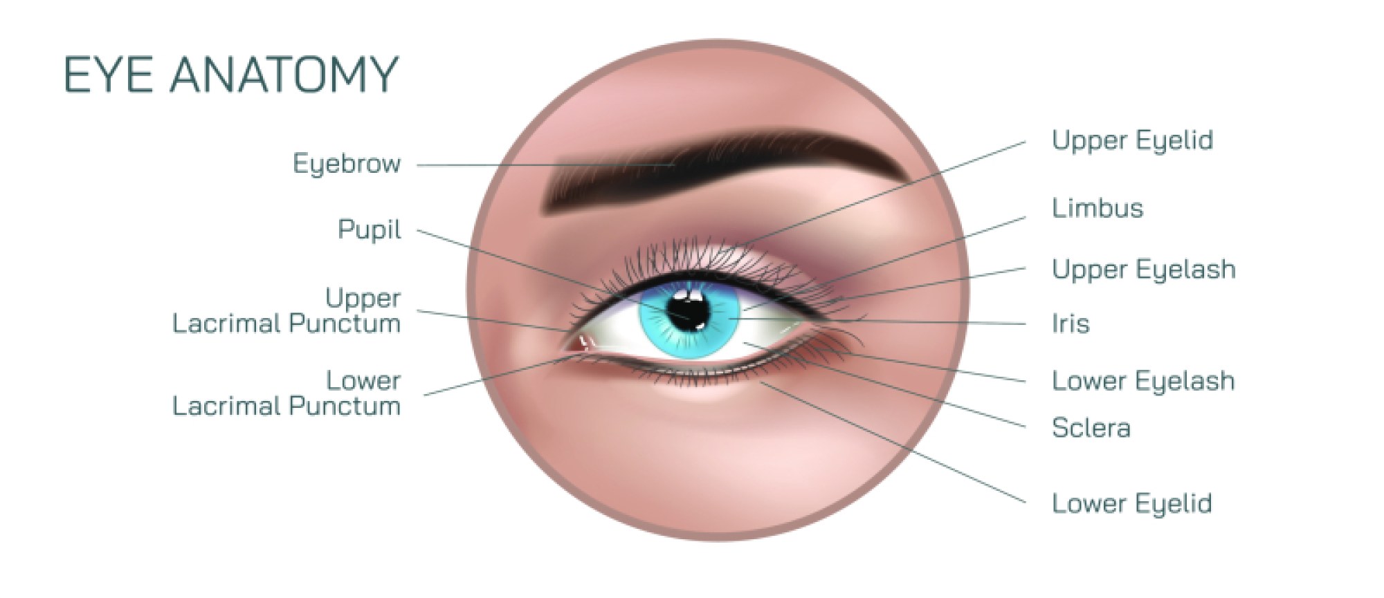
Floaters originate primarily from alterations in the vitreous body—the clear gel that fills the space between the lens and the retina. In youth, the vitreous gel is firm and uniformly transparent, maintaining a smooth, unobstructed pathway for light to pass to the retina. Over time, however, the vitreous gradually liquefies and contracts in a process called vitreous syneresis. As it degenerates, microscopic fibrils of collagen—normally evenly distributed—begin to clump together. These tiny clusters cast shadows on the retina as light passes through the eye, creating the perceptual effect of floating shapes. While the objects themselves exist inside the vitreous, the brain interprets their shadows as external shapes moving in front of the viewer. A vector illustration that depicts the eye in cross-sectional view can show this relationship clearly: clumps suspended in the vitreous, the path of incoming light, and the projection of their shadows upon the retinal surface. Such precision helps medical trainees grasp the difference between the physical location of the material and the visual phenomenon the patient perceives.
Because floaters are dynamic rather than fixed, they follow the motion of the vitreous in response to eye movement. When the eyes move quickly, floaters lag behind due to inertia; when the movement stops, they continue drifting because the vitreous gel is still in motion. A thorough medical illustration can depict motion loops and flow lines that demonstrate this inertia-based lag effect. Students studying ophthalmology benefit from understanding how this movement contributes to common patient descriptions—such as “I try to look at it but it moves away,” or “The spot stays with me no matter where I look.” The vector format is particularly valuable for this purpose because movement patterns can be layered over the static anatomy without sacrificing clarity. In animated educational modules, floaters can be shown drifting across the vitreous to give learners and patients a lifelike experience of the phenomenon.
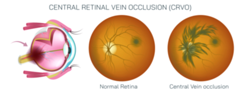
However, floaters are not uniform in cause, appearance, or clinical consequence. While age-related vitreous degeneration is the most frequent cause, floaters can also result from ocular inflammation (uveitis), trauma, hemorrhage into the vitreous cavity, complications of diabetes, or retinal tears and detachments. A comprehensive vector illustration for medical education can build awareness of this spectrum by showing multiple etiologies. For benign floaters, the graphic might show small collagen clumps suspended in liquefied vitreous. For inflammatory floaters, inflammatory cells and debris might be represented as fine granular particles. For floaters associated with vitreous hemorrhage, dark red aggregates could be shown diffusing in the gel. For floaters associated with retinal damage, the vector illustration might depict areas of traction, retinal breaks, or detachment with pigment granules spilling from the retina into the vitreous cavity. By juxtaposing normal aging changes with pathologic processes, the illustration supports clinical reasoning: not all floaters are harmless, and the context determines urgency.
One of the most important educational functions of a floaters vector illustration is to highlight warning signs that indicate possible retinal tear or detachment. While occasional mild floaters are typical with aging, sudden onset of many floaters—especially when accompanied by flashes of light or a shadow descending in the peripheral vision—can signify dangerous traction on the retina. This requires immediate ophthalmologic evaluation. Visualizing this in a diagram is far more effective than merely describing it verbally. A vector illustration can show the vitreous detaching from the retina in a posterior vitreous detachment, pulling strongly enough to tear retinal tissue, and releasing pigment cells and blood into the gel. For medical learners, this clarifies why an explosion of floaters is an ophthalmologic emergency. For patients, the visual cue reinforces the seriousness of seeking evaluation promptly to prevent permanent vision loss.
Floaters also impact visual quality beyond the simple presence of floating shapes. For some individuals, especially those who rely on precise near or intermediate focus for their profession or hobbies, floaters can significantly disrupt concentration and cause visual fatigue. When floaters drift across the line of sight repeatedly during reading, computer work, or outdoor activities in bright light, the brain becomes distracted by the constant interruption in clarity. A vector illustration that simulates how floaters distort or obscure letters, light sources, or shapes can help clinicians and caregivers empathize with patients who experience these frustrations. Although the retina remains intact, the interruption of the visual image can degrade quality of life in ways that are easy to underestimate without visual representation.
Vector illustrations are also beneficial for explaining the clinical treatment pathway. In most cases, conservative management is recommended—monitoring the floaters over time and educating patients that the brain gradually learns to ignore them through neural adaptation. For more severe or debilitating cases, treatment options may include laser vitreolysis to break up large floaters or pars plana vitrectomy to remove the vitreous gel entirely, though these procedures carry surgical risks and must be carefully selected. A vector illustration that overlays treatments onto the anatomical diagram helps clarify where interventions occur and what they aim to accomplish. The biomedical transparency of a vector graphic is particularly useful when explaining options to patients who are considering intervention for visual relief.
Diseases such as myopia and diabetes also shape the educational value of floaters illustrations. Individuals with high myopia are at greater risk of earlier vitreous degeneration and retinal detachment. Patients with diabetic retinopathy are susceptible to vitreous hemorrhage, which produces dense floaters that obscure vision. Illustrating these associations helps medical learners connect systemic diseases with ocular complications, strengthening interdisciplinary understanding across endocrinology, primary care, and ophthalmology.
Another important advantage of vector-based artwork lies in its adaptability to different learning environments. Whether a lecturer needs a full-page digital cutaway of the eye, a simplified patient-friendly version for a clinic brochure, or an animated resource for e-learning, the scalability ensures that key features remain crisp and undistorted. Labels, arrows, callouts, and progressive layers can be added without compromising the original illustration. For this reason, vector diagrams form a backbone of ophthalmology education, complementing slit-lamp photographs and imaging studies by offering standardized clarity free of physiological variances.
Floaters may seem simple on the surface—tiny shapes drifting across vision—but they involve a complex interplay of vitreous anatomy, aging, inflammatory processes, systemic disease, and retinal health. When depicted in a vector illustration, the invisible becomes understandable: the gel structure, the clumps casting shadows, the risks associated with sudden onset, and the distinctions between benign and urgent causes. For medical professionals, this visual mapping strengthens diagnostic confidence. For patients, it transforms anxiety and confusion into comprehension and proactive behavior. And for educators, it streamlines the challenge of teaching an internal ocular process that cannot be seen with the unaided eye.
In essence, a floaters in eye disease vector illustration is far more than a graphical representation. It is a tool that brings physiology, pathology, symptom experience, urgency recognition, and treatment planning into one unified visual language. By making microscopic ocular changes visible and relatable, the illustration reinforces both scientific learning and real-world protection of vision health—helping clinicians act decisively when floaters are harmless and urgently when they signal danger, ultimately supporting safer outcomes and greater understanding for every viewer it reaches.