Eye Diseases CRVO vs BRVO Vector Illustration Comparing Central and Branch Retinal Vein Occlusion with Causes, Symptoms, and Blood Flow Blockage
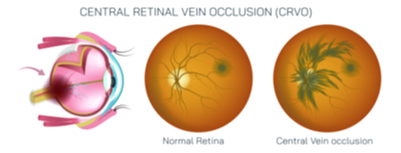
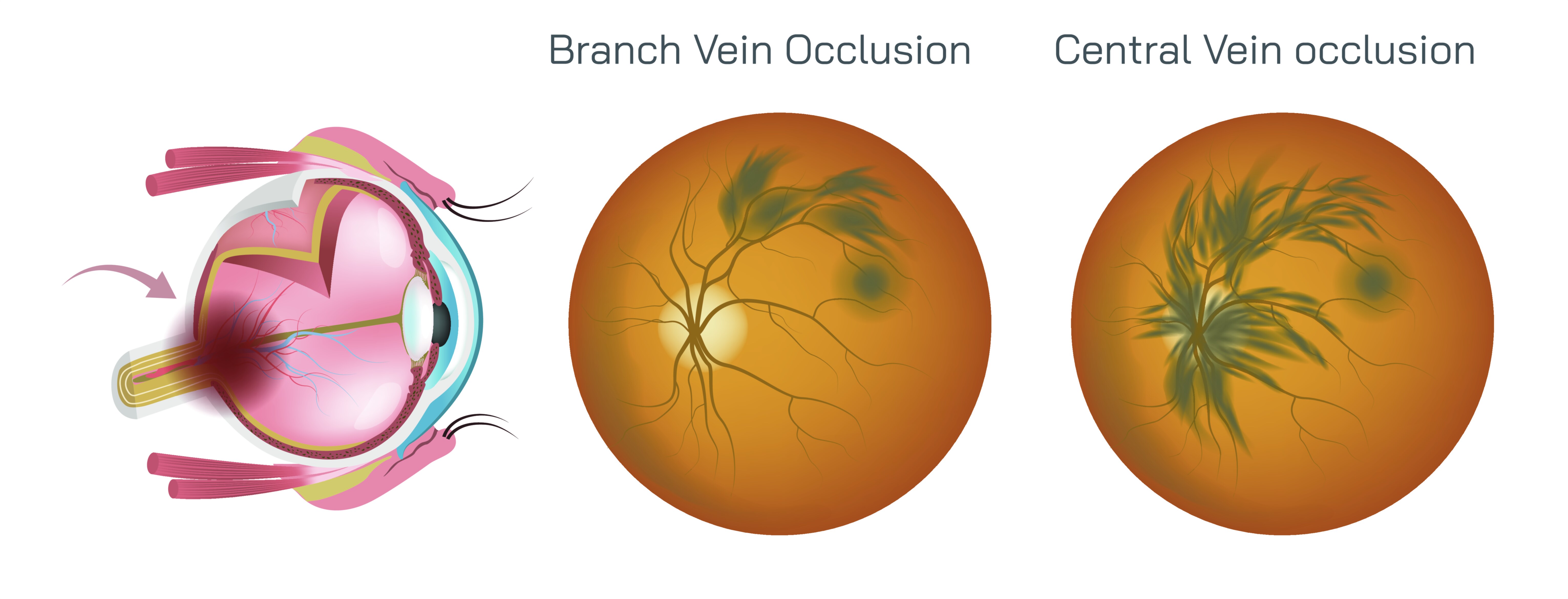
Retinal vein occlusion (RVO) is a common vascular disorder of the eye in which veins in the retina become blocked, leading to impaired blood flow, retinal hemorrhage, and vision loss. There are two primary types: Central Retinal Vein Occlusion (CRVO), which affects the main retinal vein, and Branch Retinal Vein Occlusion (BRVO), which involves one of the smaller tributary veins. A vector illustration comparing CRVO and BRVO typically integrates retinal anatomy, blood flow pathways, occlusion sites, causes, and clinical manifestations, providing a clear, educational tool for ophthalmology, optometry, and medical education. By combining labeled retinal vessels, blockage visualization, and symptomatic annotations, the diagram allows learners to understand the differences in location, severity, and visual impact between the two conditions.
At the center of the illustration is a cross-sectional or top-down view of the retina, showing the optic disc, central retinal vein, branch veins, arteries, and macula. The central retinal vein is prominently labeled, branching into smaller veins that supply specific retinal quadrants. CRVO is illustrated with a blockage at the central retinal vein near the optic disc, marked with a red or highlighted obstruction. Arrows indicate retrograde congestion and impaired blood flow throughout the retina. BRVO is depicted with a blockage in one of the branch veins, often at the crossing point of a retinal artery and vein, which is a common anatomical site of compression. Arrows highlight localized blood flow disruption confined to the territory supplied by that branch vein.
Causes of both conditions are labeled and visually integrated into the illustration. CRVO is commonly associated with hypertension, diabetes mellitus, glaucoma, and hypercoagulable states, while BRVO often occurs at arteriovenous crossings where vein compression by a thickened artery or atherosclerotic change impedes blood flow. In vector diagrams, these factors may be represented with inset symbols or icons, such as blood pressure gauges, sugar molecules, or thickened vessel walls, linking systemic risk factors to localized retinal occlusion.
The illustration also highlights blood flow changes and retinal congestion. In CRVO, arrows indicate generalized venous backup leading to retinal hemorrhages, macular edema, and optic disc swelling, illustrating the global effect on the retina. In BRVO, arrows demonstrate localized pooling of blood within a segment of the retina, often in the upper or lower quadrants, with less widespread edema compared to CRVO. Color coding differentiates oxygenated arterial flow (bright red) from deoxygenated or congested venous flow (darker red), visually reinforcing hemodynamic changes.
Symptoms and clinical signs are incorporated into vector insets. CRVO typically causes sudden, painless vision loss affecting the entire visual field, while BRVO results in partial visual field defects, corresponding to the affected retinal quadrant. Magnified insets may depict retinal hemorrhages, cotton wool spots, and fluid accumulation in the macula, labeled to show the correlation between vascular blockage and visual impairment. Arrows may indicate fluid leakage from congested veins contributing to macular edema, which is a major cause of vision reduction.
Vector illustrations often include comparative panels showing CRVO versus BRVO side by side. The CRVO panel demonstrates a blockage near the optic disc affecting all retinal veins, while the BRVO panel shows a localized branch vein obstruction with downstream effects confined to a smaller region. Labels and directional arrows illustrate differences in extent of ischemia, hemorrhage, and retinal swelling, emphasizing how the location of the occlusion determines clinical severity.
Additional features may include risk factor overlays, showing systemic conditions like hypertension or diabetes affecting vascular integrity, and arrowed diagrams of blood flow, visually indicating where circulation is impaired and how congestion propagates through the venous network. Inset magnifications may illustrate arteriovenous crossings in BRVO, showing how vein compression by overlying arteries leads to localized occlusion.
Vector diagrams may also incorporate treatment indicators such as anti-VEGF injections or laser therapy sites. Arrows and labels can indicate areas where interventions aim to reduce macular edema, restore perfusion, or prevent neovascularization, connecting anatomical pathology to clinical management. Color coding for affected versus unaffected retinal areas enhances clarity for educational purposes.
By combining retinal anatomy, central and branch vein occlusion sites, blood flow arrows, systemic risk factors, symptoms, and treatment references, a vector illustration provides a comprehensive comparison of CRVO and BRVO. It emphasizes the differences in location, extent, and impact on vision, helping learners understand how vascular blockage manifests clinically and physiologically. Color coding, directional arrows, magnified insets, and labeled structures enhance clarity and accessibility for students, clinicians, and patients.
Ultimately, a vector illustration comparing CRVO and BRVO demonstrates the relationship between retinal vascular anatomy, occlusion site, and clinical consequences, making complex ocular vascular disorders visually understandable. Through labeled veins, obstruction sites, blood flow diagrams, and symptom annotations, the diagram transforms abstract concepts of retinal vascular pathology into an educational and visually engaging framework for vision science and medical study.