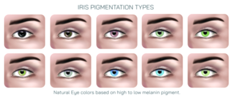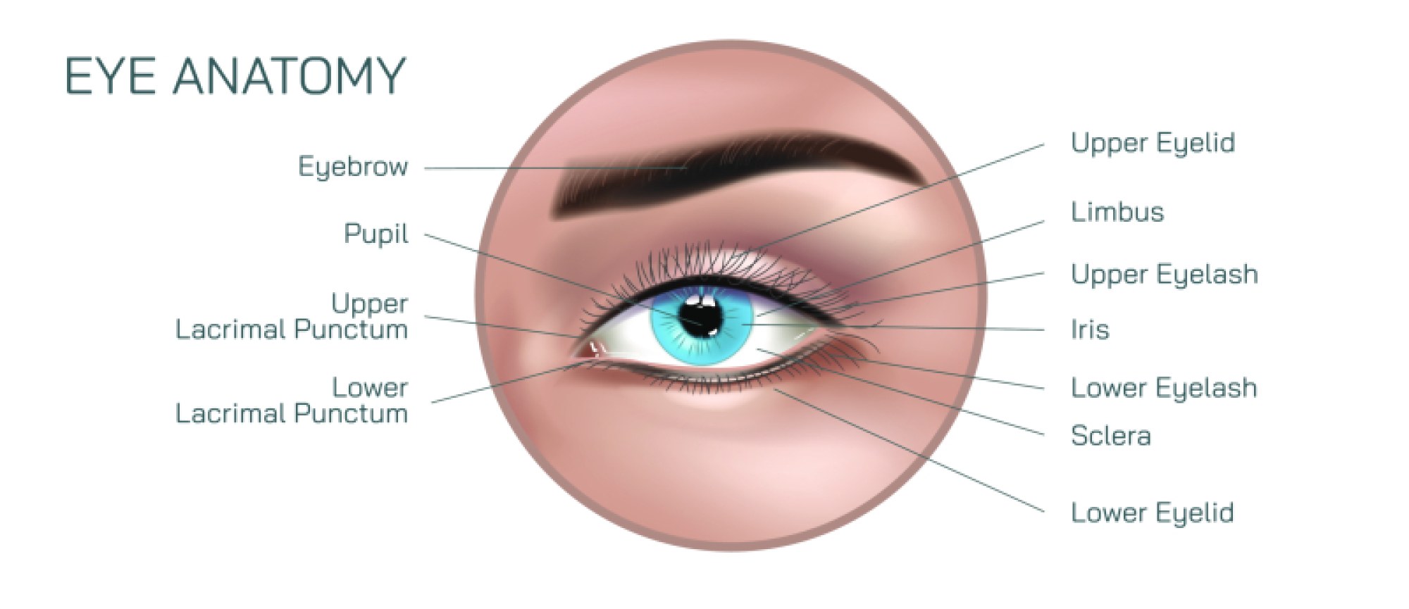
Melanin and Eye Color Showing Pigmentation Levels and Eye Color Variation
The remarkable diversity of human eye color is one of the most visually striking examples of biological variation, yet its origins lie in a microscopic pigment called melanin, distributed across specific structural layers of the iris. Although eye color is often described in simple terms such as brown, blue, green, gray, or hazel, these familiar shades are not produced by colored dyes or separate pigments unique to each hue. Rather, all natural eye colors arise from differing amounts of melanin, the type of melanin present, and the way the iris scatters and absorbs light when interacting with that pigment. An illustration or educational explanation showing melanin and eye color is therefore not merely a study in appearance, but a window into genetics, developmental biology, cellular pigmentation, and the physics of light. The iris acts not only as the muscular structure that controls pupil size but also as a canvas on which pigmentation patterns determine the familiar features of a person’s gaze. By understanding melanin levels and how they translate into eye color variation, one also comes to understand how uniquely layered biology and physics shape something as simple and recognizable as color.
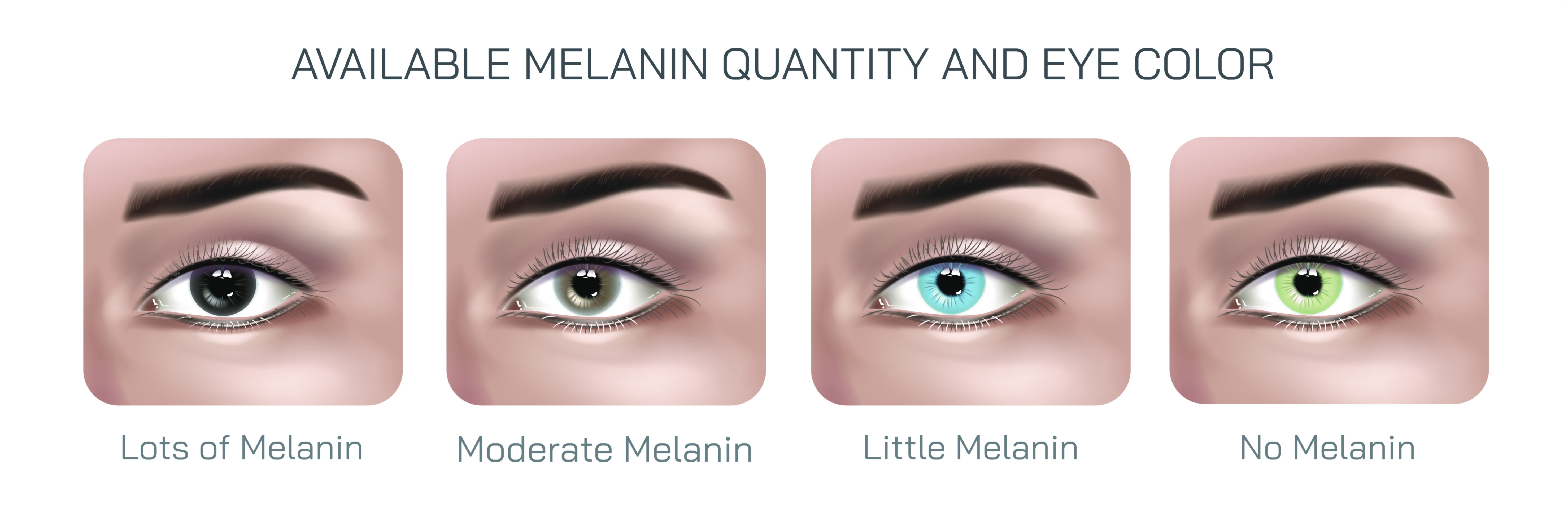
Human eyes contain two primary types of melanin: eumelanin, which is brown to black, and pheomelanin, which is reddish to yellow. These pigments are produced and stored in specialized cells known as melanocytes located within the stroma and the posterior pigment epithelium of the iris. Every iris—regardless of appearance—contains a heavily pigmented back layer known as the posterior pigment epithelium. What differentiates eye colors is the amount of pigment located in the anterior layers. Brown eyes contain abundant melanin distributed throughout the iris stroma, allowing them to absorb the majority of incoming light and reflect a deep, rich brown tone. Blue eyes, in contrast, contain very little melanin in the stroma. Because there is minimal pigment to absorb light, short wavelengths scatter in much the same way the sky appears blue. This structural scattering rather than blue pigment produces the color. Gray eyes exhibit low melanin levels as well but often have increased collagen density that diffuses light differently, leading to a cool, smoky appearance. Green eyes represent an intermediate level of melanin, where a modest presence of brown eumelanin combines with light-scattering effects to produce the greenish tone. Amber eyes, a distinct category, contain higher levels of pheomelanin, yielding a golden or coppery appearance that differs noticeably from hazel even though both contain mixed pigmentation.
Because melanin determines how much light is absorbed versus scattered, eye color is not a binary feature but a spectrum. Some irises contain a uniform pigmentation, while others present multi-tonal variation around the pupil, across the iris surface, or near the outer edge. Hazel eyes exemplify such variation, often containing gradients of brown, amber, and green tones that shift subtly depending on lighting and pupil dilation. These shades reflect uneven melanin distributions within the iris stroma, where local concentrations of pigment alter light absorption and create ring-like or radial blends of color. The beauty of these variations lies not only in aesthetics but also in insight into developmental patterns. During infancy, many children are born with blue or gray eyes due to initially low melanin activity. As melanocytes gradually increase pigment production over the first few years of life, the true eye color emerges, sometimes deepening dramatically as melanin deposits grow denser. This developmental transformation illustrates the role of gene regulation rather than simple genetic dominance.
The genetic basis of eye color is an intricate matrix rather than a single trait inherited in a predictable fashion. Early textbook models of a “brown gene” dominating over a “blue gene” have given way to a more advanced understanding of polygenic influence. Multiple genes—including OCA2 and HERC2 on chromosome 15, as well as others involved in melanin transport and production—interact to determine the final appearance of the iris. These genes control not only how much melanin is produced but how efficiently it is distributed within the iris layers. Environmental or epigenetic factors do not typically change eye color in adulthood, yet hormonal changes, certain medications, or pigment disorders may alter hue gradually over time. In rare cases, heterochromia occurs—a condition in which one eye differs in color from the other or a single iris contains two distinct pigmentation zones. This can result from genetic mosaicism, developmental variation, trauma, or medical conditions influencing the melanocytes. In such instances, eye color becomes a visual record of pigment asymmetry.
Melanin plays an additional protective role beyond color. Because its primary biological purpose is to absorb ultraviolet radiation and neutralize free radicals, darker eyes offer slightly greater natural protection against bright light and UV exposure, reflecting evolutionary adaptation to high-sunlight regions. Conversely, lighter eyes—commonly seen among populations historically native to northern latitudes—scatter more light and may be more sensitive to glare, yet do not carry a functional disadvantage in lower-UV environments. This evolutionary pattern shows how pigmentation and geography shaped one another over generations. Even though modern lifestyles, indoor work, and sunglasses have altered the practical relevance of pigmentation, the relationship between melanin and eye color still tells a population-level story of adaptation and migration.
The complexity of eye color becomes even clearer when imagining what someone actually sees through the lenses of different hues. Although people may wonder whether eye color affects visual sharpness or perception, the sensory input delivered to the brain is similar regardless of pigmentation. What changes, however, is light sensitivity. Individuals with very light eyes often report discomfort in bright sunlight or headlights at night due to reduced melanin levels that allow more internal reflection. Meanwhile, those with darker eyes may have greater resistance to glare. These experiential details add a human dimension to the biology of pigmentation, blending scientific understanding with lived experience.
In medical contexts, melanin’s influence extends beyond its role in coloration. Disorders that affect melanocyte function—such as albinism, Waardenburg syndrome, or pigmentary glaucoma—can dramatically alter iris pigmentation and ocular health. An illustration showing melanin distribution across the layers of the iris can therefore support both general anatomy instruction and specialized ophthalmology education. Eye color can also serve as a subtle diagnostic clue in clinical settings, though it is never a standalone indicator. Some inflammatory conditions may temporarily disrupt pigmentation, while long-term topical medications intended to reduce intraocular pressure can increase melanin in the iris, gradually darkening eye color. Understanding the pathways of pigment regulation helps clinicians anticipate, explain, and manage such changes.
Because melanin shapes eye color through both light absorption and scattering, the relationship between structure and shade is inseparable. The smoothness, texture, and fibrillar patterns of the iris—crypts, furrows, and radial streaks—interact with pigmentation and light to produce the final effect that the human eye perceives. Even among individuals with the same nominal eye color, no two irises are identical. Highly detailed photographic comparisons have demonstrated that each iris contains minute structural patterns that are so unique they rival fingerprints for biometric identification. This individuality is further proof that melanin distribution is not only genetically determined but sculpted through development in patterns as unique as a signature.
When examining melanin and eye color visually—from the deepest brown tones to the lightest icy gray—the diversity found within a single biological trait is astonishing. What initially seems like a simple spectrum is in reality a dynamic interplay between pigment chemistry, developmental processes, evolutionary adaptation, and optical physics. Whether observed through scientific diagrams, medical illustrations, or the simple human act of looking into another person’s eyes, eye color becomes a reminder that tiny molecular differences can produce sweeping visual diversity. It is not the presence or absence of a special pigment that defines eye color, but the degree to which melanin is present and the way it modulates light, converting invisible biological processes into visible identity.
In the end, melanin and eye color illustrate that beauty in human biology is not superficial but deeply rooted in microscopic and genetic mechanisms. Variations that seem purely aesthetic carry layers of meaning—biological history, developmental timing, evolutionary influence, and individual uniqueness. When pigmentation patterns, melanin concentration, and light-scattering behavior come together in the iris, they give each person a distinct visual signature. This convergence of science and individuality makes the study of melanin and eye color not only educational but profoundly human, reflecting the extraordinary diversity within the shared structure of the human eye.