Healthy Lungs vs Smoking Lungs Showing Normal Lung Tissue, Damaged Lungs, and Respiratory Health Comparison
The comparison between healthy lungs and smoking-affected lungs provides one of the clearest biological demonstrations of how lifestyle choices can transform internal tissue structure, organ performance, and overall respiratory health. A visual illustration of “Healthy Lungs vs Smoking Lungs” typically shows one lung with a light pink, spongy, evenly aerated texture representing normal respiratory tissue, contrasted against a darker, inflamed, congested, and structurally damaged lung that has endured prolonged exposure to cigarette smoke. While the graphic focuses on external differences in appearance, the underlying biological story is far deeper, involving cellular injury, airway obstruction, immune dysfunction, vascular damage, and progressive loss of breathing efficiency over time. The contrast between the two lungs represents not simply cosmetic change but the transition from a highly functional oxygenation system to one struggling against mechanical degradation, inflammation, and toxin accumulation.
Healthy lungs serve as the core of gas exchange, allowing oxygen from inhaled air to enter the bloodstream and enabling carbon dioxide to be removed as part of metabolic regulation. Their internal architecture is designed for maximum surface area with minimum resistance. The trachea branches into bronchi, bronchioles, and eventually millions of microscopic air sacs called alveoli, each surrounded by capillaries. In a healthy lung these alveoli remain open, elastic, and moist, allowing oxygen to dissolve across a thin membrane and pass effortlessly into circulation. The interior lining of the lungs contains cilia—tiny, hairlike structures that sweep dust, microbes, and contaminants upward toward the throat to be expelled by coughing or swallowing, keeping the airways clean without conscious effort. The tissue remains pale pink due to efficient blood flow and absence of chronic inflammation, and the elasticity of the lung walls allows effortless expansion during inhalation and smooth recoil during exhalation. In everyday life, these features translate to steady energy, good exercise tolerance, clear breathing patterns, and oxygen delivery that supports every cell in the body.
Smoking introduces a highly destructive series of events into this delicate ecosystem. Cigarette smoke contains thousands of chemicals, including tar, carbon monoxide, free radicals, heavy metals, formaldehyde, and carcinogens. When this toxic mixture reaches the lungs, the airway linings and alveolar surfaces are repeatedly irritated, inflamed, and chemically burned. In early exposure, the cilia that keep the airway clean begin to slow and eventually stop functioning. Without cilia, tar and debris accumulate along the bronchial walls, setting the stage for chronic inflammation. Over time, the lungs darken as particles from smoke and tar settle into the tissue. The illustration of “smoking lungs” often shows this visible discoloration — the once pink surface becomes mottled, brownish, or nearly black depending on exposure. These visual differences symbolize deeper damage rather than merely staining: the lungs are no longer capable of self-cleaning, oxygen transfer becomes less efficient, and chronic inflammation reshapes the architecture of the airway from the inside out.
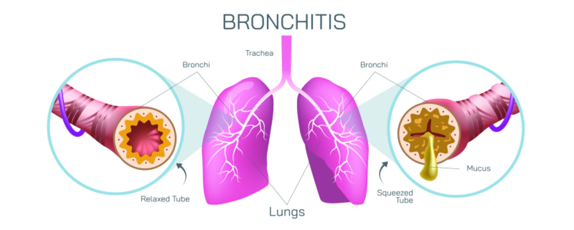
As inflammation becomes long-standing, the airways undergo structural remodeling. The bronchial tubes narrow as they swell and produce thick excess mucus. This restricted airflow leads to wheezing, shortness of breath, and persistent coughing — the familiar “smoker’s cough” that acts as the body’s last-resort attempt to clear the lungs without cilia. Repeated swelling and scarring of the bronchi set the foundation for chronic bronchitis, a component of the disease group known as chronic obstructive pulmonary disease (COPD). Alveoli also experience devastating changes under prolonged smoke exposure. Chemicals destroy their elastic walls, causing multiple alveoli to merge into enlarged, floppy sacs with far less surface area for gas exchange. This development, known as emphysema, represents one of the most serious milestones in smoking-related deterioration. In the illustration, the smoking lung appears loose, collapsed, and hollow in places because the structural scaffolding of the alveoli has been lost. As a result, oxygen may enter the lungs but cannot efficiently pass into the bloodstream, leaving the body chronically under-oxygenated and breathless even during mild activity.
The cardiovascular system suffers its own cascade of damage as smoking persists. Carbon monoxide from cigarette smoke binds to hemoglobin more readily than oxygen, reducing oxygen delivery throughout the body. The blood vessels in the lungs become damaged and thickened, restricting circulation and eventually raising pressure in the pulmonary arteries — a condition known as pulmonary hypertension. The right side of the heart must then work harder to pump blood through the lungs, increasing the risk of heart failure. The “smoking lungs” representation in illustrations does not directly show the heart, but the vascular consequences are an invisible burden that accompanies the visible tissue degradation.
On the microscopic level, smoking disrupts the immune protection that normally keeps the lungs resistant to infection. Healthy immune cells respond quickly to bacteria and viruses that enter the airway, but chronic smoke exposure weakens these cells, slows their movement, and impairs their ability to neutralize pathogens. Smokers therefore experience more frequent and more severe respiratory infections. Bacterial pneumonia, influenza complications, and persistent sinus or airway infections become much more common as lung defenses continue to degrade. The chronic inflammation also triggers an overactive immune state that begins to destroy healthy tissues in addition to damaged ones, worsening emphysema and bronchitis further. Even when a smoker is not actively sick, the lungs exist in a constant background state of inflammation, requiring more energy and effort simply to breathe.
Cancer risk is another major distinction illustrated indirectly through damaged lung tissue. Repeated exposure to carcinogens increases DNA mutation in lung cells. When enough genetic damage accumulates, cells lose their ability to regulate growth and become malignant. Tumors may form deep inside the lungs, along the bronchial tubes, or at the edges of the lung lobes. Although the “smoking lungs” image often depicts tar-darkened tissue, the darker meaning is uncontrolled cell mutation that may take months or years to become noticeable. Lung cancer is particularly dangerous because symptoms often appear late, after the tumor has already grown large or spread to other organs. The damaged appearance of the smoking lung in an illustration is therefore a visual signal of long-term cellular instability that can lead to fatal disease.
The functional consequences of lung damage steadily worsen as smoking continues. A healthy lung with full elasticity can expand deeply, allowing oxygen to fill the alveoli and keep the blood rich with oxygen during both rest and exercise. A smoking-damaged lung has lost elasticity, meaning inhalation becomes shallow and exhalation becomes incomplete. Air becomes trapped, causing the lived experience of breathlessness: the lungs feel full yet under-oxygenated at the same time. Everyday tasks such as climbing stairs, walking quickly, or even tying shoes may become exhausting. The body attempts to compensate by increasing breathing rate and heart rate, but these adjustments strain the cardiovascular system further. Meanwhile, fatigue accumulates because the brain, muscles, and organs constantly receive less oxygen than needed to function efficiently. In the most advanced stages of COPD, even speaking or eating becomes difficult because air supply is so limited.
By placing healthy lungs and smoking-affected lungs side by side, a visual illustration conveys not merely difference in color but difference in life quality. The healthy lung embodies energy, stamina, clear breathing, and strong immunity. The smoking-damaged lung reflects fatigue, shortness of breath, chronic coughing, recurring illness, inflammatory pain, restricted activity, and progressive loss of independence. The contrast continues even beyond the lungs themselves. Healthy lungs support the skin, the brain, the digestive system, and the heart because they supply continuous oxygen. Smoking-damaged lungs place strain on every system by reducing oxygen delivery and increasing toxic burden.
Yet this comparison also carries another important message: smoking damage, while serious, does not have to be irreversible if smoking stops. Once a smoker quits, cilia gradually begin to regenerate, mucus production reduces, airway inflammation decreases, and blood carbon monoxide levels normalize. While scar tissue and emphysema do not fully reverse, lung function improves, breathing becomes easier, and disease progression slows dramatically. The body continuously attempts healing as soon as the exposure ends. For this reason, the stark visual contrast between healthy and smoking-damaged lungs is not simply a warning — it is also an invitation to recovery, illustrating what the lungs can become when given the chance to heal.
In the end, the comparison between healthy lungs and smoking lungs reveals the quiet but profound influence of daily habits on the internal world of the body. One lung represents balance and effortless respiration; the other represents chronic injury and the difficulty of restoring oxygen flow against a backdrop of constant inflammation. Through structural integrity or structural collapse, through clear airways or clogged ones, through thriving alveoli or destroyed surfaces, the lungs tell the story of how well the body is supported in every breath. The illustration serves not only as a scientific explanation but as a vivid reminder of how essential healthy lungs are for movement, endurance, longevity, and quality of life — and how deeply smoking alters that story at the cellular, structural, and functional levels.