Inhale and Exhale Showing Breathing Process, Lung Expansion, and Oxygen–Carbon Dioxide Exchange in Humans
Breathing is one of the most fundamental life processes, yet its apparent simplicity hides a deeply coordinated biological mechanism that sustains every cell of the human body. The rhythmic cycle of inhalation and exhalation ensures that oxygen—an element essential for cellular energy—is continuously delivered to the bloodstream, while carbon dioxide, a metabolic waste product, is removed efficiently. The visual representation of the breathing cycle through an inhale–exhale illustration immediately captures this dual exchange: the chest expanding as air moves into the lungs and the chest relaxing as air flows out. Although the movement of air appears effortless from the outside, internally it reflects an incredibly precise orchestration of muscles, pressure gradients, lung elasticity, and gas diffusion that begins with a single breath and repeats dozens of times every minute throughout life. A closer look at this cycle reveals that breathing is not only mechanical; it is also metabolic, neurological, and continuously adaptive to the body’s needs.
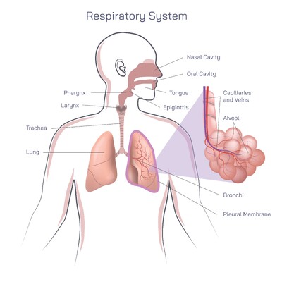
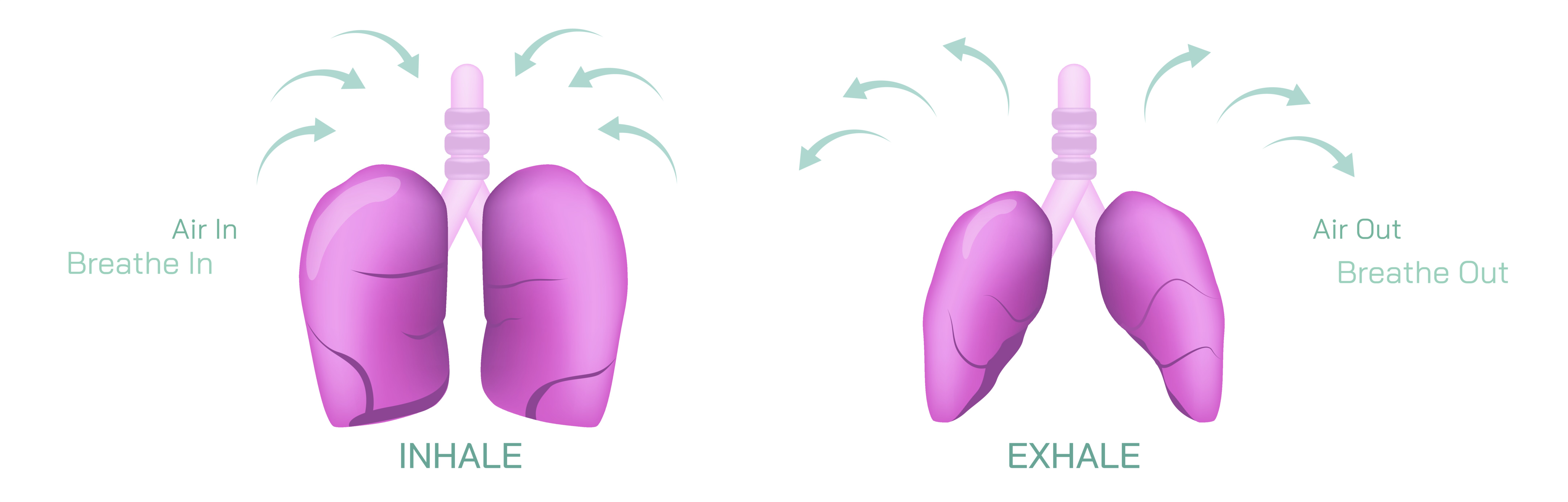
During inhalation (inspiration), the breathing process begins with the contraction of the diaphragm, a dome-shaped muscle located below the lungs. When it contracts, it flattens downward, increasing vertical space in the thoracic cavity. Simultaneously, the intercostal muscles between the ribs contract, lifting and expanding the rib cage outward. This combined movement increases the volume inside the chest, lowering internal air pressure relative to the outside atmosphere. Air flows into the lungs instinctively because gases move from higher pressure to lower pressure. As air enters through the nose or mouth, it passes down the pharynx, larynx, and trachea before reaching the branching bronchial tubes and eventually the microscopic alveoli, the tiny air sacs where gas exchange takes place. Each alveolus is surrounded by an intricate web of capillaries, forming one of the most efficient diffusion interfaces found in nature. In this phase of the illustration, the lungs appear larger and lifted, reflecting their expansion as they fill with air.
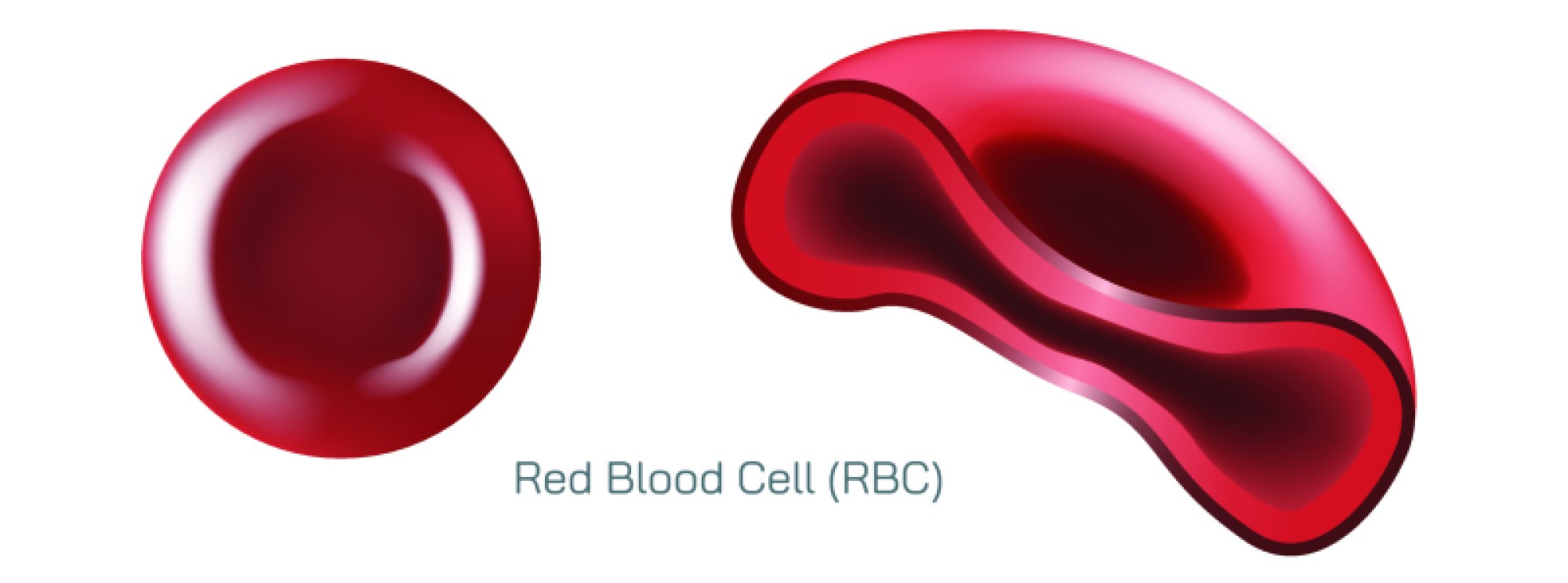
Once the incoming air reaches the alveoli, the second stage of inhalation begins—oxygen diffusion into the bloodstream. The alveolar walls are extremely thin, and the blood in surrounding capillaries has a lower concentration of oxygen than the inhaled air. This concentration gradient drives oxygen molecules to diffuse across the alveolar membrane into the plasma and red blood cells. Here, hemoglobin binds with oxygen, carrying it throughout the body via circulation. This oxygen becomes the critical fuel for cellular respiration, the biochemical process that produces ATP, the energy currency of life. Every thought, movement, heartbeat, and metabolic reaction depends on this quiet but constant transfer of oxygen with each breath. The inhale phase therefore does far more than simply fill the lungs—it sets in motion the mechanism that keeps every tissue alive.
The exhalation (expiration) process reverses the mechanics of inhalation, completing the breathing cycle. After oxygen has been delivered to tissues, carbon dioxide, produced as a waste product of cellular respiration, returns to the lungs through the bloodstream. When the concentration of carbon dioxide increases, the diaphragm and intercostal muscles relax. The diaphragm moves upward into its dome shape while the rib cage collapses slightly inward. This reduces the volume inside the chest cavity, increasing internal pressure and pushing air out of the lungs. Since the elastic tissues of the lungs naturally recoil, expiration does not require the same muscular effort that inhalation does—under resting conditions, it occurs passively. Carbon dioxide leaves the bloodstream by diffusing from the capillaries back into the alveoli, then exiting through the bronchi, trachea, and air passages before being released from the nose or mouth. In vector illustrations, exhalation is depicted as a narrowing chest and a reduced lung shape, with arrows indicating outward airflow.
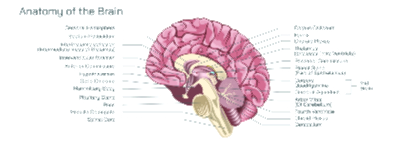
Breathing automatically adapts to changes in the body’s demands through the respiratory control centers of the brainstem, particularly the medulla oblongata and pons. These centers constantly monitor carbon dioxide concentration, blood pH, and oxygen levels, then adjust the rate and depth of breathing to maintain homeostasis. During sleep, breathing slows to conserve energy; during exercise, breathing becomes deeper and quicker to meet increased metabolic demand. Emotional states—stress, fear, excitement, and relaxation—also alter breathing patterns because the respiratory centers communicate with the limbic system and autonomic nervous system. A sigh, a gasp, or a slow deep breath becomes not only a physiological response but also an emotional signature encoded within respiratory rhythm. Thus, breathing is tightly tied to both survival and subjective experience.
The efficiency of gas exchange depends greatly on lung structure. Each lung contains roughly 300 million alveoli, creating a combined surface area roughly equal to that of a tennis court. The thinness of alveolar walls, the moist lining that promotes diffusion, and the close proximity of capillaries together create optimal conditions for oxygen and carbon dioxide transfer. Diseases that damage alveolar surfaces—such as emphysema, pneumonia, or pulmonary fibrosis—interfere with this exchange, causing shortness of breath even when airflow appears normal. Illustrations of inhalation and exhalation often highlight the alveoli to emphasize their role as the true functional core of breathing, where external air becomes biologically usable.
The breathing cycle is also central to acid–base balance, because carbon dioxide directly influences pH. When carbon dioxide builds up in the bloodstream, it lowers pH, creating acidity; when too much carbon dioxide is removed too quickly, it raises pH excessively. This balance explains why breathing changes during strenuous activity or hyperventilation and why controlled breathing techniques can calm the nervous system. Slow diaphragmatic breathing improves oxygen saturation and stabilizes carbon dioxide release, tapering stress responses by signaling safety to autonomic pathways.
From early life to the end, breathing is continuous, autonomous, and essential. Yet humans can consciously modulate it, unlike most biological functions. Techniques such as deep breathing, meditation breathing, breath-holding, and rhythmic breathing during singing or athletic training demonstrate how voluntary control can influence both mind and body. Breathing becomes a physiological link between consciousness and survival, between emotion and metabolism, and between mind and muscle.
An inhale–exhale illustration visualizes all these mechanisms at once: the expansion of the chest, the inflow of air through the respiratory passages, the filling of the lungs with oxygen, the exchange of gases at the alveoli, the return flow of carbon dioxide, and the release of air as the body resets for another cycle. While mechanically simple in appearance, the process embodies one of the most fundamental biological truths—that life is sustained through continuous exchange. Every breath renews the body, stabilizes internal chemistry, and delivers the oxygen that makes energy possible. The lungs, diaphragm, and respiratory pathways together create a rhythm that has pulsed since birth and will continue until life’s final moment. Breathing is not only an action—it is the ongoing partnership between the body and the atmosphere that makes existence possible.