Bronchoconstriction Showing Airway Narrowing, Breathing Difficulty, and Lung Function During an Asthma Attack
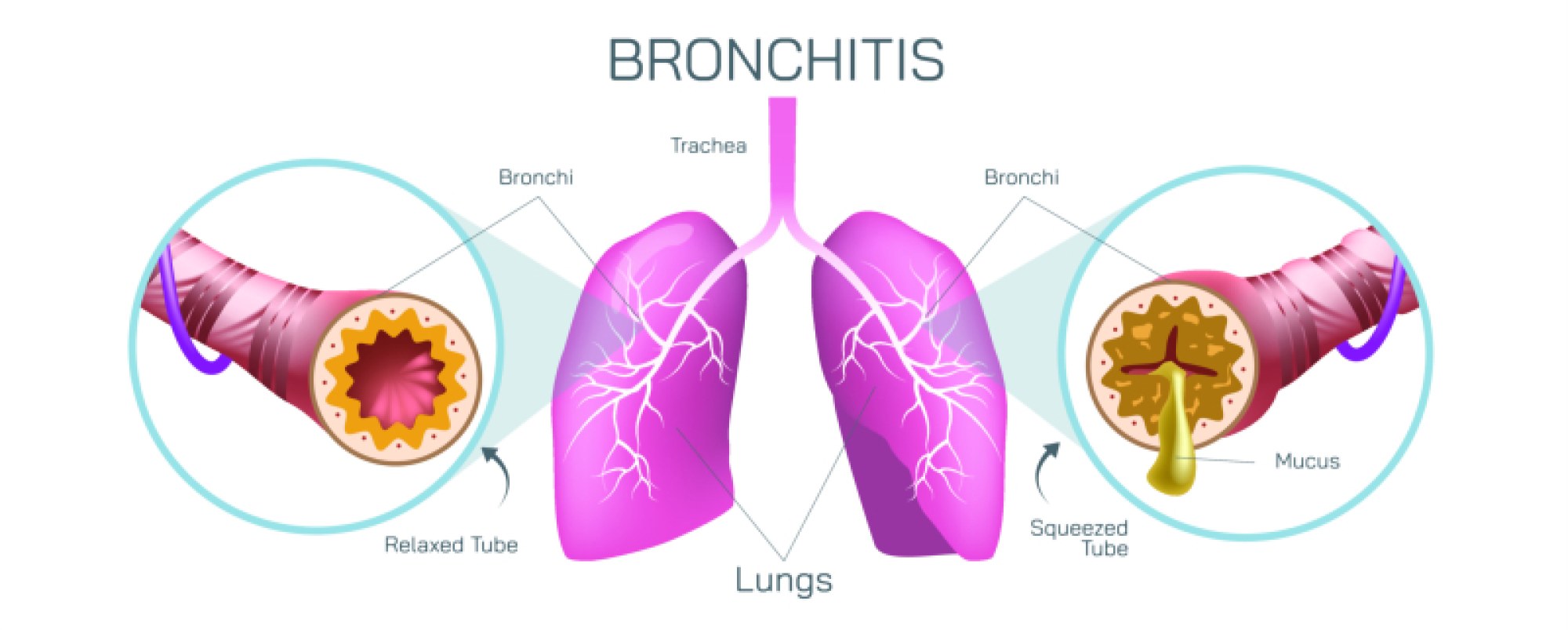
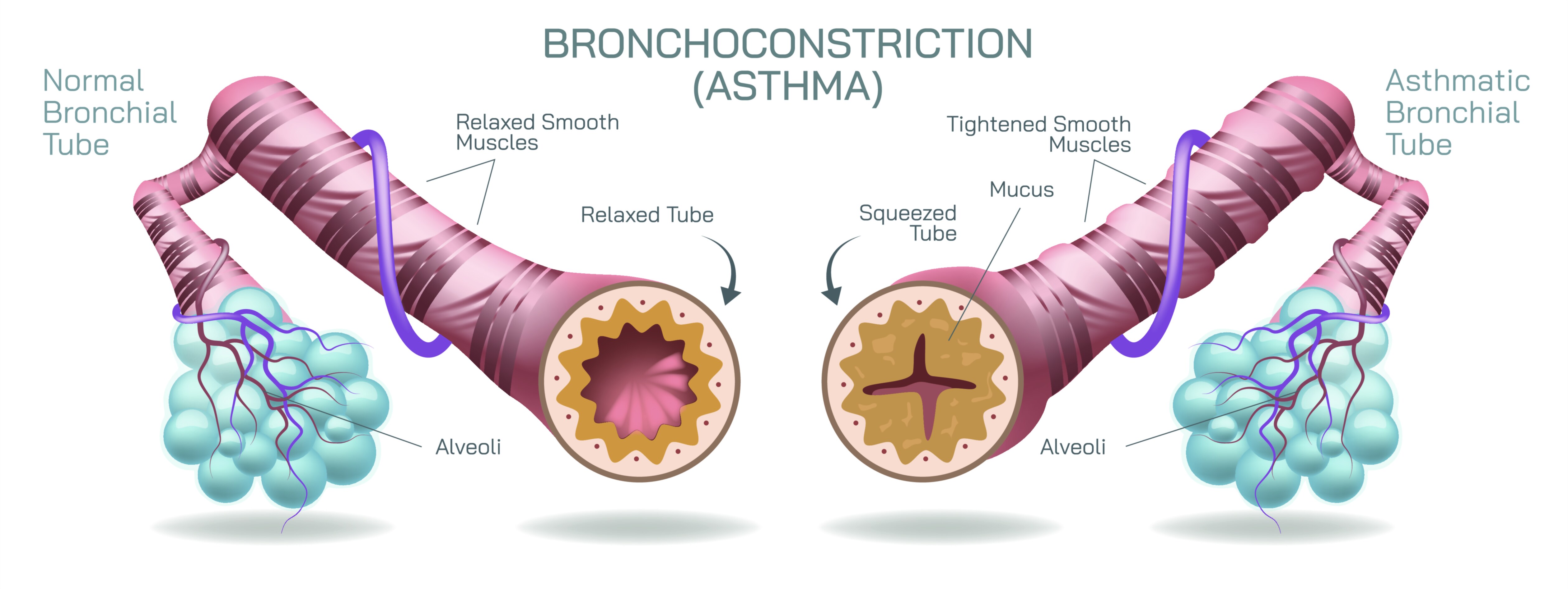
Bronchoconstriction is a defining feature of asthma and represents one of the most dramatic examples of how a small change inside the body can produce an overwhelming physical sensation. Although an illustration of bronchoconstriction may simply show narrowed airways inside the lungs, the biological reality behind this image encompasses a complex interaction between inflammation, smooth muscle contraction, and hypersensitivity of the respiratory system. Under normal breathing conditions, the bronchial tubes remain open and flexible, allowing air to move freely into and out of the lungs with minimal resistance. During an asthma attack, however, these same airways tighten sharply, the lining of the bronchial walls swells, and thick mucus fills the already restricted passages. The result is a frightening and uncomfortable experience of breathing difficulty, wheezing, chest tightness, and oxygen deprivation. A vector illustration of bronchoconstriction captures this shift visually—wide, relaxed airways suddenly becoming pinched, swollen tunnels through which air can barely pass—and helps viewers understand what makes an asthma attack so physically urgent.
To appreciate bronchoconstriction deeply, it helps to visualize the structure of the respiratory airways during a calm state. In a healthy or resting condition, the bronchi and bronchioles—the branching tubes that carry air from the trachea to the alveoli—are supported by smooth muscle bands arranged in circular rings. These muscles help regulate airway diameter, but under normal circumstances they remain relaxed. The airway lining, called the mucosa, sits flat and uncongested, producing only a thin layer of mucus to trap dust and pathogens. Even during exercise or rapid breathing, the bronchioles expand slightly to allow faster airflow, ensuring that oxygen supply meets the body’s increasing metabolic demands. When represented visually, this calm airway appears as a spacious passage with smooth, evenly shaped walls and a clear opening through which air flows effortlessly.
Bronchoconstriction disrupts this equilibrium suddenly and intensely. When asthma triggers activate the immune system—whether allergens, viruses, cold air, smoke, stress, exercise, pollution, fragrances, or occupational irritants—the tissues inside the airway respond as though defending the body from threat. Immune cells release chemical mediators such as histamine, leukotrienes, and cytokines, which rapidly cause the smooth muscle rings around the airways to tighten. Because the bronchial tubes are soft and flexible, this muscular contraction reduces their diameter like tightening a drawstring. At the same time, inflammatory swelling thickens the airway walls, while mucus glands shift into overproduction, filling the narrowed tubes with sticky secretions. In an illustration of bronchoconstriction, the lumen—the central opening of the airway—shrinks dramatically, sometimes until it appears only as a tiny slit surrounded by inflamed tissues.
This narrowing has immediate consequences for lung function. During inhalation, the diaphragm attempts to draw air downward into the lungs, but because the bronchioles have constricted, incoming air meets resistance. The person must work harder to bring each breath in, often lifting their shoulders or using neck muscles in an effort to expand the chest. Yet it is exhalation that becomes even more difficult. Air becomes trapped inside the lungs because it cannot flow smoothly back out through the narrow passages. This trapped air creates a sensation of tightness and fullness that many people describe as “trying to breathe through a straw” or “having a weight on the chest.” The body reacts to this obstruction by increasing breathing rate, but faster breathing does not solve the obstruction; it can intensify fatigue and anxiety, worsening the cycle.
The hallmark sound of an asthma attack—wheezing—comes from air squeezing turbulently through narrowed airways. Yet one of the most concerning stages of bronchoconstriction occurs when wheezing suddenly disappears. This does not signify improvement but indicates that airflow has become so restricted that barely any air is moving—an emergency that requires immediate intervention. In severe bronchoconstriction, the person may struggle to speak, feel panicked or exhausted, develop bluish lips or fingernails due to low oxygen, or experience altered consciousness. These symptoms reveal how narrowing within the bronchioles impacts not just the lungs but the entire body.
Inside the alveoli, the final destination of every inhaled breath, gas exchange depends on airflow reaching these tiny air sacs. When bronchoconstriction prevents enough fresh oxygen from entering the alveoli, oxygen levels in the blood drop. Meanwhile, carbon dioxide, normally exhaled easily, accumulates in the bloodstream, upsetting the body’s delicate acid–base balance and threatening organ function. The brain senses this change and sends urgent signals to breathe harder and faster. In a vector illustration, alveoli may appear normal while the airway above them is severely constricted, demonstrating that the problem is not in the air sacs themselves but in the passages that feed them. This reinforces why asthma medications focus primarily on relaxing airway muscles and reducing inflammation rather than on the alveoli directly.
Bronchoconstriction arises from a unique characteristic of the asthmatic airway—hyperresponsiveness. In people with asthma, the bronchi and bronchioles react excessively to triggers that would barely affect someone without the condition. The smooth muscles contract more strongly than necessary, and inflammatory cells stay primed for exaggerated responses, creating a pattern of chronic sensitivity. Over time, recurring inflammation can lead to airway remodeling, in which the walls of the bronchioles become permanently thickened and less elastic. This long-term change can make asthma symptoms more frequent or severe. Illustrations of bronchoconstriction often add a developmental layer by showing how recurrent attacks—even if reversible—can gradually alter the structural architecture of the airways.
Despite its intensity, bronchoconstriction is reversible with appropriate treatment. Medications known as bronchodilators, delivered through inhalers or nebulizers, target the smooth muscle bands and relax them within minutes, widening the airway and improving airflow. Anti-inflammatory medications such as corticosteroids reduce swelling and mucus production, preventing future attacks and restoring airway sensitivity to a healthier baseline. Spacers, peak flow monitoring, allergy management, trigger avoidance, and controlled breathing techniques all support long-term stability. A visual diagram of bronchoconstriction paired with a comparison to a medicated, reopened airway powerfully illustrates the relief that follows effective treatment.
Breathing difficulty during asthma is not simply a matter of airflow; it is the embodiment of a deep biological response designed for protection that becomes maladaptive in asthma. Bronchoconstriction transforms a calm, open airway into a compressed tube struggling to sustain life-sustaining gas exchange. It engages muscular, inflammatory, immune, and neurological pathways simultaneously, and its effects ripple far beyond the lungs—from oxygen supply to cognition, comfort, and emotional regulation. A vector illustration showing bronchoconstriction helps viewers grasp these complexities instantly: wide bronchioles becoming narrow, mucosa swelling, mucus filling the space, and arrows depicting obstructed airflow.
More than a medical diagram, such an illustration becomes a window into the lived experience of asthma. It explains why a crisis can escalate rapidly, why early intervention is essential, and why asthma should be treated as a chronic inflammatory disorder—not merely a breathing inconvenience. Through the imagery of airway narrowing and lung function disruption, bronchoconstriction reveals the immense importance of open airways to human life and the delicate balance that every breath depends on.