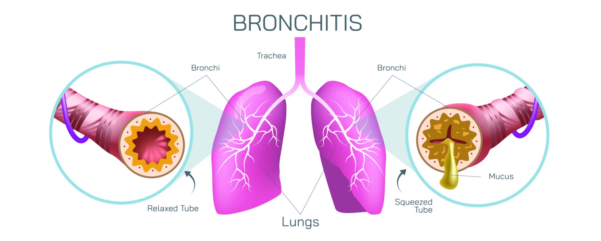
Bronchitis Illustration: Inflammation of Bronchi and Respiratory Tract Explained
Bronchitis is a respiratory condition centered around inflammation of the bronchi—the large and medium-sized airways that deliver air to and from the lungs—and a detailed illustration of this disorder visually communicates how much disruption a single region of the respiratory system can cause. In a healthy respiratory tract, the bronchi form smooth, cylindrical tubes lined with a thin, moist mucous membrane and fine hair-like structures called cilia. These cilia move rhythmically to sweep inhaled particles, dust, and pathogens upward toward the throat, keeping the airways clear and protecting the delicate lung tissue. The inner walls of the bronchi appear pink, open, and unobstructed, providing free airflow during breathing. A cross-section illustration of a healthy bronchus shows a wide circular airway, minimal mucus, and an organized ciliary surface working together to maintain efficient ventilation.
In bronchitis, this peaceful anatomical scene changes dramatically. An illustration of an inflamed bronchus highlights swelling of the mucous membrane causing thickening of the airway wall. The once spacious airway narrows considerably, reducing space for air to pass in and out. The cilia, overwhelmed by inflammation, become clogged or paralyzed, and their sweeping motion slows or stops entirely. Meanwhile, the mucus-producing glands inside the bronchi respond to irritation by secreting excessive mucus. In a cross-section view, this mucus appears as thick layers or plugs coating the bronchial lining, further blocking airflow. The airway no longer functions as an open breathing channel but instead becomes constricted, sensitive, and congested. This structural illustration helps explain why individuals with bronchitis experience persistent coughing—the body is struggling to expel trapped mucus—and why breathing can feel tight or labored.
Illustrations of bronchitis often divide the condition into acute and chronic forms, each of which presents structural differences visible within the bronchi. Acute bronchitis develops quickly, often following a viral infection of the upper respiratory tract such as a cold or flu. In this version, the bronchial lining becomes temporarily inflamed and swollen, with exaggerated mucus secretion. In a visual representation, the inflammation in acute bronchitis may appear bright red, with thick mucus occupying a significant portion of the airway. The inflammation gradually reduces as the infection clears, and the airway returns to normal as the cilia recover. Chronic bronchitis, however, appears much more dramatic in long-term illustrations. Prolonged irritation—commonly from cigarette smoke, pollution, or chemical inhalation—causes persistent swelling and structural remodeling of the bronchi. The airway walls become thickened and scarred over time, the cilia are sparse or permanently damaged, and the mucus glands enlarge and overproduce secretion. In a detailed illustration, the airway cross-section becomes severely narrowed and obstructed, and mucus fills the passage chronically rather than temporarily. Chronic bronchitis often forms part of COPD (Chronic Obstructive Pulmonary Disease), and comparing its bronchial structure with that of acute bronchitis and a normal airway makes the progression of damage visually unmistakable.
Airways affected by bronchitis also become unusually reactive, meaning they are highly sensitive to environmental triggers. In inflammation-focused illustrations, the bronchial muscle layer encircling the airway appears thickened and tense, sometimes partially constricting in response to irritants or cold air. This reactivity causes temporary spasms—bronchospasm—that tighten the airway even more and cause wheezing or shortness of breath. Many illustrations show these spasms by drawing narrowed muscular rings squeezing the inflamed airway like tightening bands. For some people, especially those with asthma, bronchitis can trigger prolonged episodes of airway hyperreactivity that significantly limit airflow.
One of the most instructive visual features of bronchitis illustrations is the comparison between normal mucus clearance and impaired mucus clearance. In a healthy airway, cilia are shown upright and rhythmic, sweeping particles upward along a thin layer of mucus. In bronchitis, cilia appear flattened and matted under thick mucus that sits stagnant instead of flowing outward. This visual depiction explains not only coughing but the sensation of mucus “stuck in the chest” that patients often describe. It also highlights why hydration, warm air, and expectorants help: thinning mucus allows cilia to move it again. When the mucus is thick, it becomes fertile ground for bacterial growth, which is why acute viral bronchitis can sometimes evolve into secondary bacterial infection.
Bronchitis illustrations also highlight how inflammation affects oxygen exchange indirectly. The bronchi themselves are not responsible for absorbing oxygen—that happens in the alveoli deep within the lungs. But when airflow into the lungs becomes restricted due to narrow, mucus-filled bronchi, less air reaches the alveoli for gas exchange. Medical illustrations often pair images of the bronchi with simplified alveoli shown beyond them to demonstrate that blocked airways may leave many alveoli under-ventilated. This mismatch between air supply and the body’s oxygen demand makes breathing shallow, inefficient, and exhausting. A diagram showing increased effort of breathing—using accessory muscles of the chest and neck—helps explain why even mild bronchitis can cause fatigue and why chronic bronchitis forces the lungs and heart to work harder over time.
Environmental factors are also commonly represented visually because they play such a large role in both triggering and preventing bronchitis. Cigarette smoke exposure may be illustrated as particulate matter entering the airways, overwhelming cilia, and triggering inflammatory reactions. Pollution, dust, workplace chemicals, and respiratory viruses are likewise shown as tiny irritants that latch onto the bronchial lining, setting off cascades of inflammation. Some diagrams highlight protective behaviors by contrasting a polluted and smoke-filled airway with a clean and well-ventilated one. These illustrations communicate that bronchitis is not simply an internal process; it is profoundly shaped by air quality and respiratory hygiene.
Healing stages of bronchitis can also be represented visually. In acute bronchitis, as inflammation dwindles, the lining gradually regains its smoothness, cilia recover upright orientation, and mucus production returns to normal. In contrast, healing from chronic bronchitis may show only partial recovery because structural changes such as scarring and gland enlargement make the airway permanently narrower. Illustrations comparing reversible and irreversible airway changes convey why chronic bronchitis demands long-term management and prevention rather than short episodic treatment.
Ultimately, a bronchitis illustration is more than a cross-section of an inflamed airway—it is a biological story about how irritation transforms respiratory stability into discomfort, obstruction, and impaired airflow. It explains the origins of persistent cough, wheezing, chest tightness, and breathlessness while showing how mucus, inflammation, airway reactivity, and ciliary damage interact. It also places respiratory care in context: hydration, rest, avoidance of irritants, and when necessary, medications work not only because they relieve symptoms, but because they promote the restoration of a clear, open airway that can once again deliver air effortlessly to the lungs. Through visual representation of swollen bronchi beside healthy ones, the mechanisms of bronchitis become easier to understand, allowing the condition to be seen not merely as coughing but as a temporary—or in some cases chronic—disruption of one of the body’s most vital pathways for sustaining life.