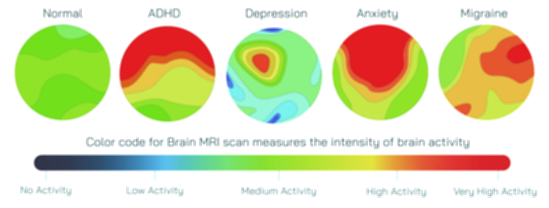
MRI Scan and Color Code Showing Brain Imaging, Medical Diagnosis, and Anatomical Mapping Visualization
Magnetic Resonance Imaging, widely known as MRI, represents one of the most transformative advancements in modern medical diagnostics, allowing physicians and neuroscientists to visualize the human brain with extraordinary precision without requiring invasive procedures or exposure to harmful radiation. An MRI scan does more than create a simple picture of brain tissues; it captures the internal structures, water content, and molecular movement inside the brain, converting subtle physiological signals into detailed anatomical maps. When these images are enhanced with color coding, the scanning process becomes an even more powerful form of visualization, translating grayscale data into maps that reveal regional brain function, abnormal tissue, nerve pathways, blood flow, and neurochemical activity. The result is a multidimensional view of the brain that supports diagnosis, research, treatment planning, and an ever-expanding understanding of how brain anatomy is linked to behavior, cognition, and disease.
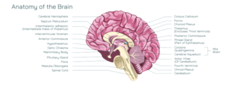
An MRI scan is based on the interaction between magnetic fields, radiofrequency waves, and hydrogen atoms within the body. Because the brain contains a high amount of water and lipids, its tissues respond clearly to these electromagnetic signals. During scanning, hydrogen protons align inside a strong magnetic field, then release measurable signals when stimulated by controlled pulses. Sophisticated algorithms transform these signals into layered images that represent slices of the brain from various directions—axial, coronal, and sagittal. When these layers are stacked together, they form a 3D representation of brain anatomy. Every fold and groove of the cerebral cortex, every deep structure such as the thalamus or hippocampus, every white matter tract connecting distant regions can be captured without physically touching the brain. MRI therefore provides not just a picture of where things are, but a method for revealing how the internal brain landscape changes with development, injury, disease, or age.
Standard MRI scans typically appear in shades of black, white, and gray, with different tissue types displaying unique contrast patterns. Gray matter, white matter, cerebrospinal fluid, and vascular spaces each respond differently to magnetic and radio signals because of their density and molecular composition. This helps radiologists detect tumors, bleeding, inflammation, lesions, structural deformities, and signs of degenerative disorders such as multiple sclerosis, Alzheimer’s disease, or Parkinsonian changes. However, grayscale alone cannot communicate the full complexity of brain structure or function, and this limitation is what motivated the integration of color coding into MRI visualization. When color is applied, the same raw imaging data gains a clarity that the human eye processes more intuitively.
Color-coded MRI does not simply tint the image; it represents dynamic information mapped onto the anatomy. In structural imaging, color can highlight tissue composition differences so distinctly that subtle abnormalities become easier to detect. In diffusion tensor imaging (DTI), color maps depict the direction of white matter fiber tracts, revealing the communication highways that connect brain regions. A single map may use red to signify left–right neural pathways, blue to mark vertical pathways, and green to represent front–back connections. This mapping illuminates how the brain’s networks are organized and how injuries such as traumatic brain injury or stroke disrupt them. In functional MRI (fMRI), color shows brain activity in real time by detecting oxygen level changes in blood flow. Warm hues such as yellow and red represent areas of high activity, while cooler tones such as blue or purple show less activation. Through this layering, clinicians can observe which parts of the brain respond during speech, memory tasks, emotional processing, pain response, or problem-solving.
Color-coded MRI visualization transforms diagnosis into a deeper understanding of brain function. For neurological disorders, it distinguishes between tissue that is structurally intact but functionally impaired and tissue that is physically damaged. In psychiatric disorders, altered activity patterns can be mapped to regions responsible for emotion, decision-making, or reward, offering biological insight into conditions once understood only through behavior. In epilepsy, color-coded scans help pinpoint the seizure onset zone, guiding surgeons with accuracy that protects vital functional areas. For brain tumors, multi-contrast and multi-color MRI differentiate tumor boundaries, edema, necrotic tissue, and healthy tissue, supporting safer surgical removal and more targeted treatment. In developmental scans, color mapping illustrates how children’s brains form pathways, how adolescents refine emotional and cognitive circuits, and how aging rewires connectivity over time.
Beyond clinical medicine, color-coded MRI contributes enormously to research and education. It allows scientists to visualize how brain networks light up when experiencing music, solving mathematics, interacting with others, or recalling memories. It reveals how meditation modifies attention networks, how learning strengthens synaptic pathways, and how lack of sleep disrupts default mode networks. The ability to translate invisible neural patterns into visible maps bridges the gap between biological processes and subjective experience. Students, patients, and scientists alike can comprehend brain function more intuitively because color gives shape to processes that would otherwise remain abstract.
The value of MRI becomes even more evident when combined with technology that tracks disease progression over time. A sequence of color-coded scans can show how a treatment shrinks a tumor, how physical therapy restores connectivity after stroke, or how a neurodegenerative disorder slowly alters anatomy. This longitudinal perspective enables truly personalized medicine because it reveals not just the presence of disease but the pace and pattern of change. Every pixel of the scan becomes a point of reference, contributing to a comprehensive understanding of the brain’s evolution.
Even the act of interpreting a color-coded MRI carries emotional impact. When patients see the areas of their brain associated with memory, speech, movement, and personality highlighted in vivid detail, they gain a visual understanding of their condition. For many, this fosters hope rather than fear because it transforms the brain from a mysterious organ into a readable map. Treatment becomes a guided journey rather than an abstraction, grounded in data that is clear and personal.
Ultimately, MRI and color-coded visualization show how medicine is not merely about detecting disease, but about revealing the structural and functional symphony of the brain. It enables diagnosis with striking clarity, guides treatment with precision, educates through visual communication, and deepens scientific inquiry into how the mind arises from neural tissue. Every scan holds a story: the architecture of thought, the signature of memory, the imprint of emotion, and the silent traces of injury or healing. Through MRI color coding, the brain becomes both mapped and understood, its hidden processes transformed into vivid representations that strengthen the connection between biology and human experience.