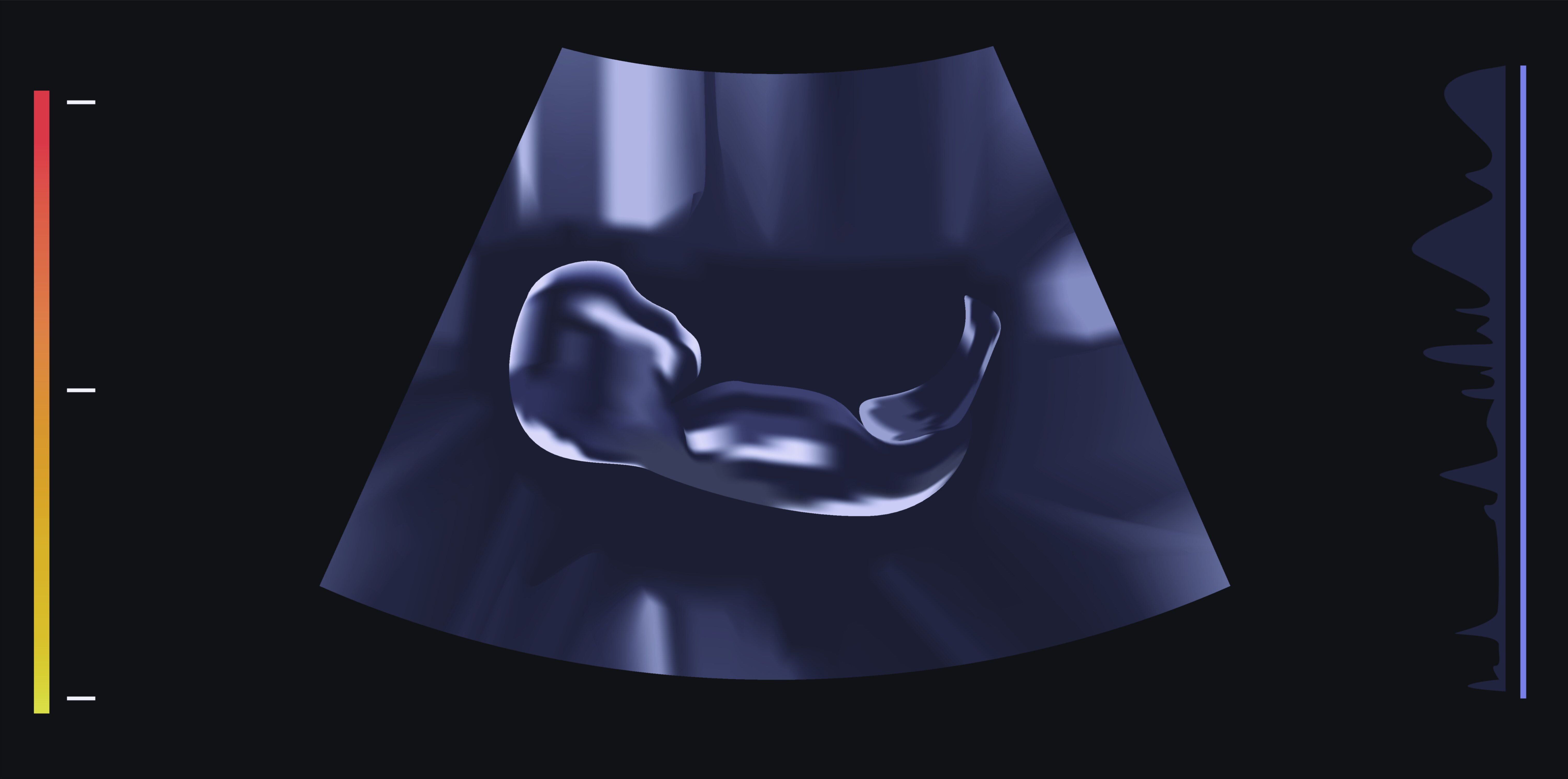
Ultrasound Image — Medical Sonography Scan
Ultrasound imaging, also known as medical sonography, represents one of the most transformative and widely used diagnostic tools in modern healthcare, offering a unique way to visualize internal structures of the human body without exposing patients to ionizing radiation or invasive procedures. At its core, an ultrasound image is formed by high-frequency sound waves that penetrate the body, reflect off tissues, and return to a transducer, where the signals are converted into real-time visual representations. Although the principle sounds straightforward—project sound into the body and capture the echoes—the behavior of these waves inside biological tissues, their interactions with organs, and the sophisticated processing required to interpret these signals give rise to an extremely rich and versatile imaging method. The resulting images offer a window into the body’s structure and motion, revealing anatomical details, blood flow patterns, organ health, and fetal development with remarkable clarity and safety. Ultrasound imaging thrives at the intersection of physics, biology, technology, and clinical expertise, making it both scientifically intricate and profoundly human in its role within patient care.
The fundamental principle behind ultrasound imaging begins with the use of sound waves that are far above the range of human hearing. These waves are generated by a handheld device called a transducer, which uses piezoelectric crystals to convert electrical energy into mechanical vibrations. When pressed against the skin, sometimes aided by a conductive gel to improve acoustic coupling, the transducer sends these high-frequency waves into the body. As the sound waves travel through tissues, they encounter boundaries between structures with different densities and acoustic properties. Each boundary reflects a portion of the wave back toward the transducer. This returning energy is detected by the same crystals, now acting in reverse to convert mechanical echoes back into electrical signals. These signals undergo rapid processing by the ultrasound machine, which interprets their strength and timing to construct an image. Bright or white areas on the ultrasound correspond to strong echoes from dense or highly reflective structures, such as bones or connective tissue, while darker or black regions indicate areas where sound passes through easily, such as fluid-filled structures.
What makes ultrasound images especially compelling is their real-time nature. Unlike static imaging modalities, ultrasound provides continuous live feedback from inside the body, allowing clinicians to observe the movement of organs, the beating of a fetal heart, the flow of blood through vessels, or the dynamic contraction of muscles and tendons. This real-time capability makes ultrasound ideal for guiding medical procedures, from biopsies to fluid drainage to needle placement. A practitioner can watch the needle’s progress on-screen, ensuring precision and reducing risk. This interactive quality distinguishes ultrasound from many other imaging techniques and makes it indispensable across emergency medicine, obstetrics, cardiology, musculoskeletal assessment, and diagnostic radiology.
Ultrasound’s versatility extends deeply into patient care, beginning with its most iconic application: prenatal imaging. Sonography allows clinicians and expectant parents to visualize developing fetuses safely and repeatedly, monitoring growth, detecting abnormalities, assessing gestational age, and observing fetal movement. These scans provide invaluable insight into the well-being of the pregnancy without any harm to the mother or child. But ultrasound reaches far beyond obstetrics. In cardiology, echocardiography uses ultrasound to examine the structure and function of the heart, capturing images of valves, chambers, and blood flow patterns. Because the heart is constantly moving, ultrasound’s real-time imaging is uniquely suited to evaluating cardiac performance, diagnosing heart diseases, measuring ejection fraction, and detecting abnormalities such as valve dysfunction or congenital defects. In the abdominal region, ultrasound can assess the liver, gallbladder, kidneys, pancreas, spleen, and major blood vessels, revealing conditions such as gallstones, liver disease, cysts, tumors, or aneurysms. Its ability to differentiate between fluid-filled and solid structures makes it particularly effective in investigating abdominal pain, swelling, or abnormal laboratory findings.
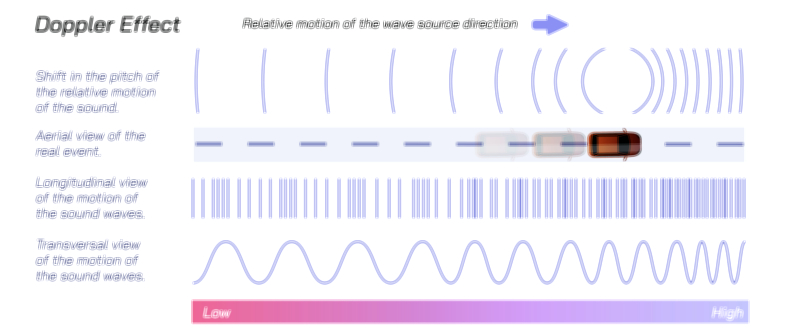
Musculoskeletal ultrasound provides another window into the body, offering high-resolution imaging of tendons, ligaments, joints, and soft tissues. Unlike MRI or X-ray, ultrasound allows examination during movement, making it ideal for diagnosing sports injuries, tendon tears, joint inflammation, and fluid accumulations. In vascular imaging, Doppler ultrasound enhances the basic imaging mode by measuring the motion of blood cells within vessels. This technique helps clinicians evaluate blood flow direction and speed, detect blockages, diagnose deep vein thrombosis, assess arterial narrowing, and monitor circulation in grafts or stents. Doppler imaging contributes significantly to stroke prevention and cardiovascular assessment, showing how ultrasound evolves beyond anatomical visualization into functional analysis.
One of the hallmark strengths of ultrasound lies in its safety profile. Because it uses sound waves instead of ionizing radiation, it poses no radiation-related risks to patients, allowing frequent use in sensitive populations such as pregnant individuals or children. This safety advantage is paired with portability and accessibility. Many ultrasound machines are compact and wheeled, while handheld devices connect to tablets or smartphones, making ultrasound one of the few imaging modalities that can be brought directly to the bedside, ambulance, operating room, athletic field, or remote medical clinic. This mobility democratizes diagnostic imaging, allowing prompt assessment even in environments lacking advanced equipment. The absence of a need for large infrastructure or specialized shielding means ultrasound is widely available, cost-effective, and practical for routine evaluations as well as emergency diagnostics.
Despite its advantages, ultrasound imaging has limitations that arise from the physics of sound wave behavior. Sound waves do not travel well through air or gas, making lungs and gas-filled intestines difficult to image. Bone reflects nearly all ultrasound energy, preventing visualization of structures behind bony surfaces, such as the brain in adults. Image quality can also vary based on the skill of the operator, as sonography requires careful probe positioning, angle control, and anatomical knowledge. Certain body types, including patients with a high degree of soft tissue thickness, can attenuate sound waves, reducing clarity. These limitations highlight the need for a trained and experienced practitioner, who must not only produce high-quality images but also interpret subtle variations in echo patterns. Unlike automated imaging modalities, ultrasound interpretation is partly an art, shaped by experience, observational sensitivity, and familiarity with normal and abnormal tissue appearances.
Nonetheless, ultrasound continues to evolve through technological advancements. Modern machines incorporate 3D and 4D imaging, providing volumetric representations and dynamic motion views that enhance fetal visualization, surgical planning, and structural assessment. Elastography, an advanced ultrasound technique, evaluates tissue stiffness to help diagnose conditions like liver fibrosis or thyroid disease. Contrast-enhanced ultrasound uses special agents to improve visualization of blood flow in organs and tumors, expanding diagnostic accuracy. High-resolution transducers offer unparalleled detail in superficial structures, while improved Doppler capabilities refine vascular assessments. These innovations push ultrasound closer to the sophisticated precision of other imaging modalities while maintaining its low-risk, accessible nature.
In clinical practice, the ultrasound image serves as far more than a picture; it is an interpretive tool shaped by the interaction of physics, anatomy, and clinical reasoning. The grayscale patterns, shadow formations, enhancement zones, and echo textures form a visual language that practitioners learn to read with fluency. For example, fluid appears dark because it allows sound to pass freely, whereas dense tissues like bone create bright echoes with deep shadows behind them. Cysts, solid tumors, blood clots, and inflamed tissues all produce distinctive echo signatures. Recognizing subtle differences in these patterns enables early detection of disease, targeted treatment decisions, and continuous monitoring of progress or recovery.
Ultimately, ultrasound imaging stands as a remarkable fusion of sound and sight, transforming invisible internal vibrations into meaningful diagnostic information. It bridges the gap between patient and clinician by providing immediate visual insight without invasive procedures. Whether it is used to guide a surgeon’s hand, evaluate a newborn’s organs, diagnose a potentially life-threatening condition, or reveal the first glimpse of a developing life, ultrasound imaging holds a profound and enduring role in medical practice. It embodies the principle that technology can illuminate the hidden workings of the human body in ways that are safe, immediate, and deeply informative, making sonography one of the most essential pillars of modern diagnostic medicine.