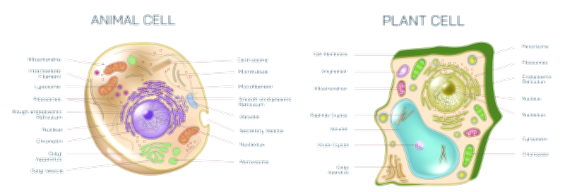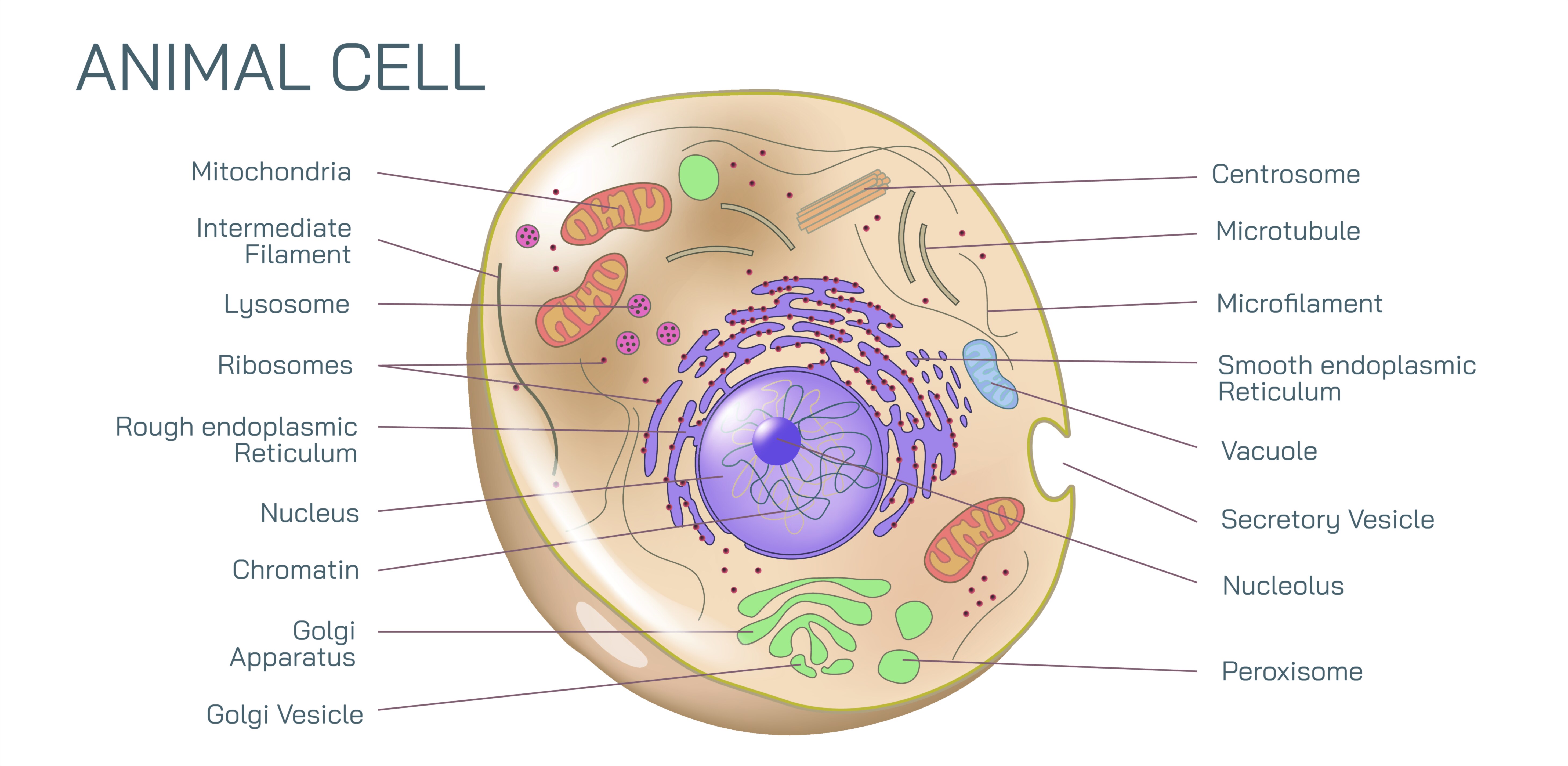
Animal Cell Showing Organelles, Nucleus, Cytoplasm, and Internal Structure of the Eukaryotic Animal Cell
The eukaryotic animal cell is one of the most remarkable biological structures in nature, functioning as a self-contained living system that sustains growth, communication, repair, energy production, and genetic continuity. When viewed under a microscope or illustrated in a biological diagram, the interior of an animal cell reveals a highly organized world where every component contributes to survival in its own specialized way. Unlike simple prokaryotic cells that lack internal compartmentalization, the animal cell contains numerous membrane-bound organelles, each with its own biochemical environment and function. These organelles divide cell operations into coordinated tasks — energy processing, protein synthesis, waste breakdown, nutrient storage, molecular transport, and genetic regulation — ensuring that even the smallest physiological action depends on exquisite structural precision. The image of an animal cell therefore represents more than shape and symmetry; it is a visual map of life at the microscopic scale.
At the center of animal cell structure lies the nucleus, a defining feature of eukaryotic life. The nucleus acts as the command and control center of the cell because it houses DNA, the long double-stranded molecule encoding all the genetic instructions needed for growth, repair, metabolism, and reproduction. These DNA molecules are wrapped around proteins called histones and organized into chromatin, allowing enormous stretches of genetic code to fit efficiently into the confined space of the nucleus. When the cell prepares to divide, the chromatin condenses into visible chromosomes to ensure accurate genetic distribution to daughter cells. The nucleus is surrounded by a double nuclear envelope — a membrane containing pores that regulate the flow of molecules in and out — allowing selective exchange of RNA, ribosomal components, and regulatory proteins without compromising genetic safety. Deep within the nucleus sits the nucleolus, where ribosomal RNA is synthesized and ribosome subunits begin assembly before being exported into the cytoplasm. The nucleus stands as the guardian of heredity and the orchestrator of cellular activity, anchoring the identity of the cell through every moment of its existence.
Surrounding the nucleus lies the cytoplasm, a semi-fluid gel-like medium that fills the interior of the cell. Although often depicted simply as empty space in diagrams, the cytoplasm is a highly dynamic matrix composed of water, dissolved ions, enzymes, and structural filaments that support biochemical reactions. Countless metabolic processes take place here — breakdown of nutrients, assembly of molecules, signaling events, and structural rearrangements during movement or division. Suspended throughout the cytoplasm are the organelles, each enclosed within membranes that define their individual roles while allowing them to communicate and exchange components. The cytoplasm also contains the cytoskeleton, a network of microtubules, microfilaments, and intermediate filaments that provide shape, strength, and mobility. Through constant remodeling, the cytoskeleton enables processes such as intracellular transport, muscle contraction, and changes in cell shape during development, repair, or immune response.
Among the most essential cytoplasmic structures are the mitochondria, the powerhouses of the cell. These double-membrane organelles generate cellular energy in the form of ATP through aerobic respiration. Their inner membrane forms folded cristae that maximize surface area for energy-producing reactions, while their own circular DNA and ribosomes reflect an evolutionary origin rooted in ancient symbiosis. Cells with high energy demands — such as muscle, nerve, and cardiac cells — contain large numbers of mitochondria, underscoring the direct relationship between organelle specialization and functional needs. In addition to ATP production, mitochondria play roles in metabolism, heat generation, and cell death signaling pathways, positioning them as essential regulators of cellular life cycles.
Another major feature of the animal cell is the endoplasmic reticulum (ER), which exists in two continuous types: rough ER and smooth ER. The rough ER, covered in ribosomes, is the primary site of protein synthesis for proteins destined for membranes, secretion, or lysosomes. As ribosomes build proteins, these molecules are folded and modified within the ER’s interior channels before being transported to the next stage of processing. The smooth ER lacks ribosomes but plays a vital role in lipid synthesis, detoxification of harmful substances, and regulation of calcium ions, especially in muscle and liver cells. Together, the ER forms an interconnected factory where materials for cellular use and export are manufactured and prepared.
From the ER, materials travel to the Golgi apparatus, a stack of flattened membrane sacs often compared to a packaging and distribution center. Here, newly synthesized proteins and lipids are further modified — sugars may be added, molecular tags may be attached, and folding may be finalized to ensure biological functionality. Once processed, these molecules are sorted into vesicles that transport them to their final destinations: the cell membrane, secretory pathways, lysosomes, or storage compartments. The Golgi apparatus therefore acts as a transit hub that maintains order within the molecular traffic of the cell.
For cellular recycling and defense against molecular damage, the cell depends on lysosomes, which contain powerful digestive enzymes housed within acidic compartments. Lysosomes break down damaged organelles, engulfed pathogens, and unusable macromolecules, turning molecular waste into reusable raw materials. This process of controlled breakdown not only prevents toxic buildup but also supports cellular longevity by continuously refreshing internal components. Working alongside lysosomes, peroxisomes detoxify harmful byproducts of metabolism and break down fatty acids, adding another layer of internal protection against oxidative stress.
The cell membrane, or plasma membrane, forms the outer boundary of the animal cell and contributes to identity, communication, and control. Composed of a phospholipid bilayer embedded with proteins, receptors, cholesterol, and carbohydrate chains, the membrane separates the internal environment from the external world while remaining selectively permeable. Nutrients, ions, and signaling molecules are continuously regulated through channels, pumps, and transport vesicles. Receptor proteins allow the cell to detect hormones, neurotransmitters, and environmental cues, meaning that even an external stimulus triggers responses only when recognized at the membrane level. The membrane also participates in cell-to-cell recognition and adhesion, ensuring tissue cohesion in multicellular organisms.
Many diagrams of animal cells also highlight structures critical for cell division and intracellular organization. Centrioles, located within the centrosome region, help organize microtubules and form the spindle fibers that draw chromosomes apart during mitosis. Without this mechanism, genetic material would not be distributed accurately, placing genetic integrity at risk. Some cell types include specialized organelles for their functions — for example, secretory cells contain expanded Golgi structures for high protein export, immune cells contain amplified lysosomal activity for pathogen destruction, and muscle cells are packed with mitochondria to meet energy demand. This adaptability demonstrates that although one diagram may represent the “standard” animal cell, real cells vary enormously depending on the roles they perform in the body.
Every organelle is essential, but what makes the animal cell extraordinary is the coordination among its parts. The nucleus turns genetic instructions into RNA messages; ribosomes translate those messages into proteins; the ER and Golgi system refine and distribute the products; mitochondria fuel the entire operation; lysosomes remove waste; the cytoskeleton organizes shape and motion; the membrane orchestrates communication across the cellular boundary. Each part works in harmony with the others — and when even one organelle becomes dysfunctional, the consequences ripple throughout the entire cell, reflecting the interconnectedness of cellular systems.
To visualize an animal cell is to appreciate the biological infrastructure that supports life on the smallest meaningful scale. It contains structure and motion, storage and synthesis, communication and adaptation. It maintains identity while continuously exchanging matter and signals with its surroundings. It adapts to challenges through molecular intelligence encoded in DNA and executed through thousands of coordinated chemical reactions every second. The internal structure of the animal cell stands as one of nature’s greatest engineering achievements — complex yet perfectly ordered, fragile yet resilient, microscopic yet foundational to every tissue, organ, and system in the human body.