𝐏𝐥𝐚𝐭𝐞𝐥𝐞𝐭𝐬 𝐢𝐧 𝐌𝐢𝐜𝐫𝐨𝐬𝐜𝐨𝐩𝐞 𝐁𝐥𝐨𝐨𝐝 𝐂𝐞𝐥𝐥 𝐎𝐛𝐬𝐞𝐫𝐯𝐚𝐭𝐢𝐨𝐧
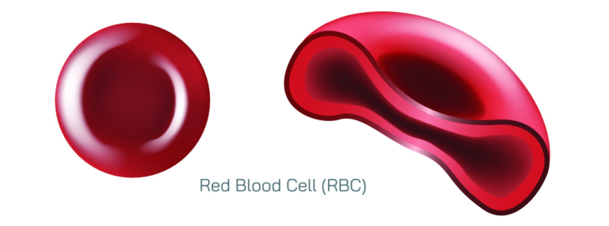
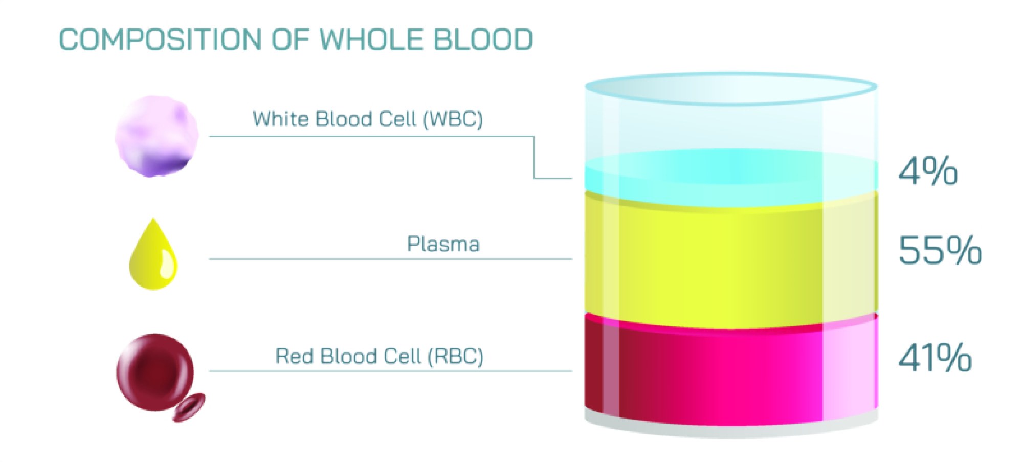
Platelets are one of the most fascinating components of human blood when observed under a microscope because of their distinctive appearance, their crucial biological function, and their ability to reflect a person’s internal health status through their quantity, size, structure, and behaviour. Blood is a complex connective tissue composed of cells suspended in plasma, and unlike red blood cells and white blood cells, platelets do not exist as complete cells but rather as small, irregularly shaped fragments of larger bone marrow cells called megakaryocytes. When examined microscopically on a stained blood smear, platelets appear as tiny purple-violet to blue granulated structures scattered among larger red cells, usually concentrated in uneven clusters rather than uniformly distributed. Their fragmented shape and small size make them easy to distinguish visually from erythrocytes, which appear as smooth, round, and biconcave structures, and from leukocytes, which are significantly larger and contain visible nuclei. This distinct morphology enables laboratory experts to analyse platelet count, size variations, clumping behaviour, structural irregularities, and staining characteristics to diagnose various blood disorders, clotting problems, and systemic diseases affecting the haematological system.
When platelets are viewed under a microscope, the first observation that stands out is their size relative to other blood cells. A typical platelet is about one-third the diameter of an average red blood cell. Instead of a smooth surface like erythrocytes, platelets show a granular or speckled texture due to the presence of alpha granules and dense granules inside them, which store clotting proteins, growth factors, calcium, and chemical messengers necessary for healing and immune response. In a healthy blood smear, platelets appear in moderate numbers, usually scattered rather than concentrated in large groups, and their sizes remain relatively consistent with minimal variation. The staining procedure often used, such as Wright or Giemsa stain, helps highlight their internal components, making them appear slightly darker at the centre and lighter towards the edges. Under high magnification, some platelets appear round while others look slightly spiky or irregular due to natural shape changes that prepare them for clotting, indicating that platelets are not static fragments but dynamic structures constantly ready for activation and repair.
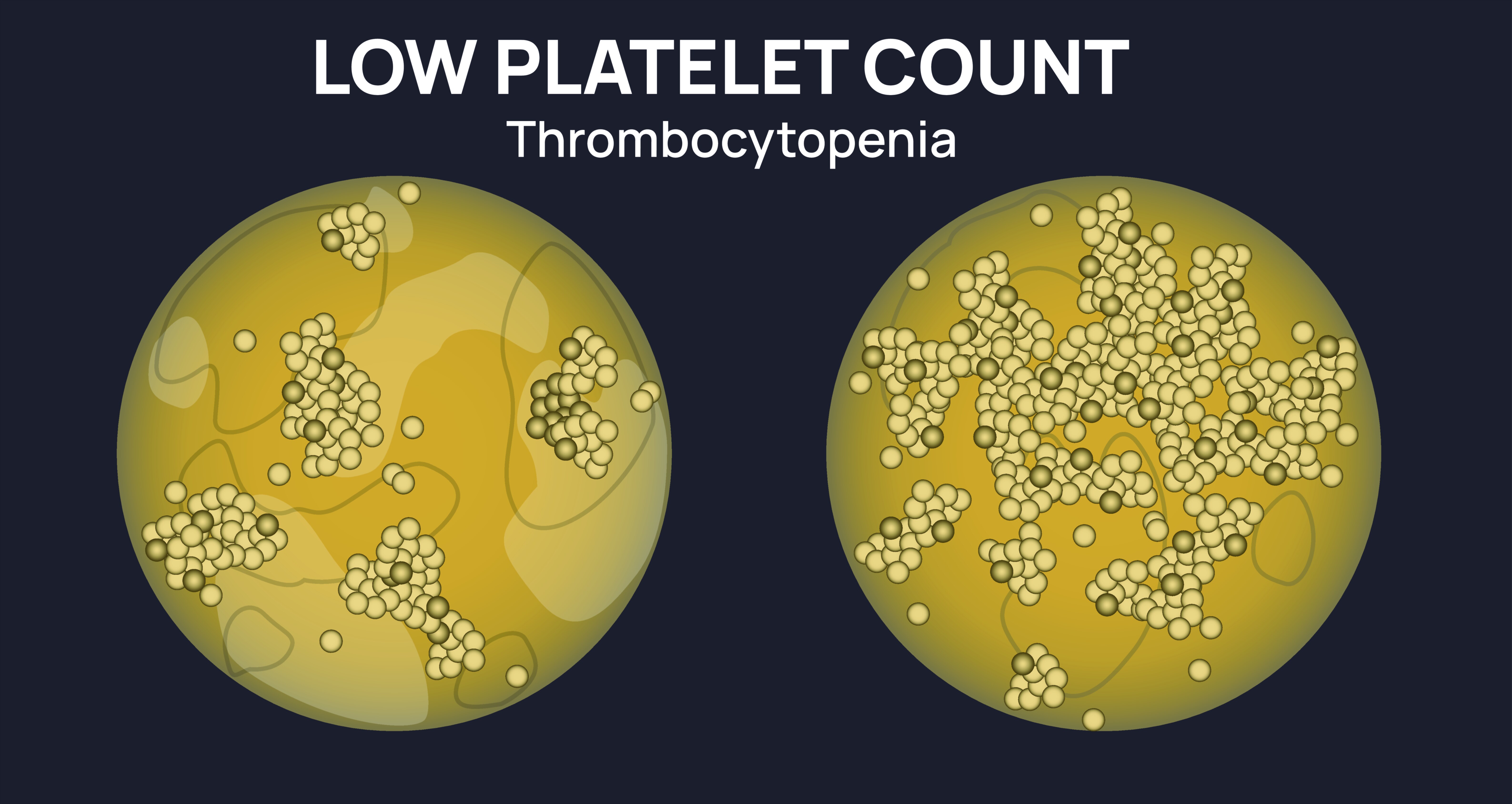
One of the most important observations when analysing platelets under a microscope is the presence or absence of clumping. Platelet clumps are clusters of many platelets sticking together, which occur when platelets activate too easily or when blood collection techniques trigger premature aggregation. In a peripheral smear, clumps may be seen near the edges of the smear and can lead to falsely low platelet counts in automated tests because the machine interprets clumps as a single particle instead of multiple platelets. Clumping can indicate disorders of excessive activation, immune responses, certain medications, and blood sample errors. On the other hand, a smear with almost no platelets or very few of them scattered irregularly may indicate thrombocytopenia, a condition in which platelet numbers are abnormally low. A smear that shows unusually large platelets, even as large as red blood cells, can indicate disorders of platelet production, genetic platelet diseases, or increased turnover where the body rapidly produces immature platelets to compensate for destruction. Irregular shapes, reduced granulation, or pale staining may signal nutritional deficiencies, bone marrow failure, or systemic disease.
Beyond simple counting and morphology, platelet observation under a microscope provides deep insight into the body’s clotting mechanisms and healing capacity. When a blood vessel is injured, platelets are the first responders that rapidly rush to the site, adhere to the exposed tissue, and initiate clot formation to prevent blood loss. The granules stored inside platelets release substances that recruit additional platelets, activate clotting factors, and help form a stable fibrin network that seals the wound. The microscopic behaviours of platelets reflect the readiness of the body to respond to injury; for example, activated platelets appear spiky or star-shaped with long extensions called pseudopods, whereas resting platelets appear round or oval with smooth edges. Observing these activation patterns helps clinicians understand platelet reactivity levels, and in disorders such as thrombocytosis or hypercoagulability, many platelets may appear activated even without tissue damage. Conversely, in bleeding disorders such as von Willebrand disease or immune thrombocytopenia, platelets may be present in normal numbers but fail to show proper granule distribution or activation morphology.
Microscope-based platelet evaluation not only supports diagnosis but also strengthens laboratory correlation with clinical symptoms. A patient with frequent nosebleeds, gum bleeding, or easy bruising may show a smear with low platelet count, abnormally large platelets, or defective granulation patterns that confirm impaired clotting response. Similarly, individuals with chronic inflammatory disorders or cancers may show elevated platelet counts with variations in size that reflect heightened bone marrow activity. Certain infections such as dengue fever dramatically reduce platelet numbers and can be monitored by observing smears repeatedly during recovery. Nutritional deficiencies, especially deficiencies of vitamin B12 and folic acid, produce large, poorly functioning platelets, while iron deficiency may produce smaller ones. Microscopic platelet evaluation also assists in detecting bone marrow failure, drug-induced platelet suppression, hypersplenism, and autoimmune platelet destruction, making it an invaluable tool in modern haematology.
The beauty of platelet observation lies in the way these tiny structures reveal the dynamic balance between coagulation, immunity, tissue repair, and inflammation. Although invisible to the naked eye, platelets orchestrate complex healing processes essential for survival. The microscope transforms them from abstract biological terminology into visible evidence of the extraordinary activity taking place within the bloodstream every second. A single smear can tell a story about the body’s internal state, whether healing, fighting disease, or struggling with deficiency or imbalance. For medical students, laboratory professionals, and clinical researchers, platelet morphology provides a real-time window into the functioning of the circulatory system, amplifying the importance of careful sample preparation, staining, and interpretation.
Understanding platelets through microscopic analysis deepens appreciation for the blood’s complexity and the precision of biological regulation. They remind us that even the smallest cellular fragments can hold immense physiological power. Each platelet carries the ability to stop bleeding, build tissue, resist infection, and maintain vascular health. Their observable patterns reflect a sensitive equilibrium that keeps the body functioning smoothly. Whether increasing in number in response to inflammation or decreasing dangerously due to immune imbalance or infection, platelets visually narrate the ongoing dialogue between injury and repair. Observing and studying them enriches scientific knowledge and medical practice, strengthening diagnostic accuracy and patient care. By placing these minute, vibrant structures on a microscope slide, the unseen processes of life become visible, connecting clinical science with the fascinating microscopic world circulating silently within each human being.