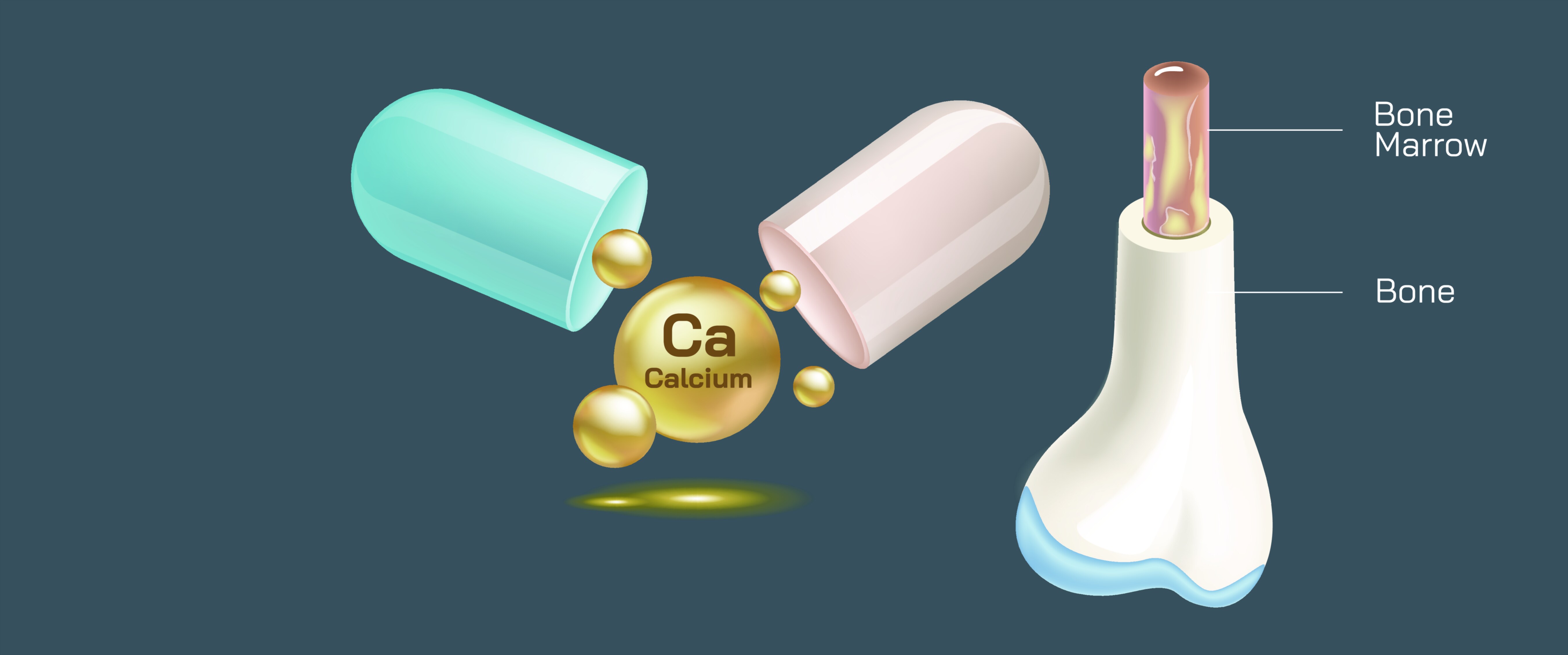
Bone and Bone Marrow Showing Calcium Importance for Strength and Healthy Structure
The human skeleton is far more than a framework for movement and body shape; it is a living organ system that protects vital tissues, stores essential minerals, produces blood, and supports metabolic stability every hour of life. A vector illustration showing bone and bone marrow with an emphasis on the role of calcium offers a visually intuitive explanation of how skeletal health is continuously built and preserved. Although most people associate calcium with strong bones, fewer understand the complex story behind how bones store, release, and reorganize calcium to maintain both structural hardness and internal body function. The combination of bone architecture and marrow activity forms a symbiotic system in which physical stability and blood production coexist. By highlighting the microscopic and macroscopic processes at play—mineral deposition, marrow activity, and nutrient exchange—a visual representation helps viewers see that calcium’s importance goes beyond density; it supports the lifelong balance between bone strength, cellular health, and the maintenance of life-sustaining functions.
In every skeletal bone from head to toe, calcium forms the mineral backbone that allows bones to withstand physical impact. Bones derive their strength from a natural composite of hard minerals and flexible collagen fibers. Collagen establishes the framework of bone tissue, while calcium and phosphorus crystallize into hydroxyapatite, filling that framework with compressed hardness. Without these crystals, bones would bend and fracture under pressure. When an illustration portrays dense mineralized bone next to an unmineralized version, the difference is striking: the healthy bone shows thick layers of compact tissue, tightly arranged trabecular struts, and solid surfaces adapted to bear weight. The undermineralized bone, in contrast, shows porous spaces, softened structure, and weak resistance to force—an immediate visual reminder of why calcium is essential and why its deficiency gradually weakens posture, mobility, and long-term skeletal function. What makes calcium unique among nutrients is not only its contribution to bone density but the way bones depend on its constant presence. Calcium does not simply enter bone once; it is continuously borrowed and restored throughout life.
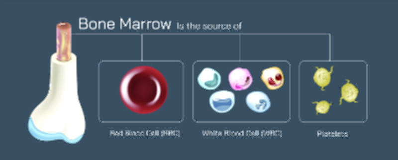
Nested within the interior cavity of many bones lies bone marrow, an essential organ for blood formation. Red marrow produces red blood cells that deliver oxygen, white blood cells that protect against infection, and platelets that aid clotting. Yellow marrow stores fats that support metabolic needs and can convert back into red marrow in times of crisis. A vector representation of marrow activity brings visibility to processes that are normally invisible. It shows how stem cells gradually develop into mature blood cells and how circulation draws these cells into the bloodstream through a network of sinusoidal channels. Because calcium-rich bones provide the cradle for this activity, skeletal health directly influences immune function, energy distribution, and the body’s ability to heal wounds and recover from injury. Healthy marrow does not function in isolation; it relies on a structurally strong and nutrient-supported bone environment to maintain cell production. A visual pairing of dense outer bone and active inner marrow helps demonstrate that the skeleton is essential to both mechanical strength and physiological survival.
Calcium plays a critical metabolic role that extends beyond the skeleton, and this is where the illustration becomes especially educational. The body relies on calcium not only for bone rigidity but also for muscle contraction, heart rhythm regulation, blood clotting, cell signaling, and nerve transmission. Because these functions are indispensable, the body maintains tight control over calcium levels in the bloodstream. When dietary calcium is insufficient, the body retrieves calcium from the bones to keep the blood supply stable. A vector depiction of calcium moving from bone into blood demonstrates how bones act as reservoirs rather than static structures. This system protects life in emergencies but damages skeletal strength over time. When calcium withdrawal happens repeatedly without adequate replenishment, bones become gradually undermineralized even if outward symptoms remain unnoticed. An illustration showing the progressive thinning of trabecular bone and the erosion of cortical mass emphasizes how silent calcium deficiency can be, often going undetected until fractures or posture changes emerge. The message becomes clear: calcium intake must match calcium requirements continuously, not intermittently, to protect bone integrity across decades.
The distribution of healthy bone structure changes with age, and calcium availability influences this process at every stage. In childhood and adolescence, rapid skeletal growth demands abundant calcium, vitamin D, and protein to build dense mass that will form the body’s long-term bone reserve. A vector graphic that displays robust, thick trabeculae in youth visually explains why calcium intake during this period is critical for establishing peak bone mass. In adulthood, calcium helps maintain equilibrium between bone breakdown and rebuilding. Bones still remodel continuously, but calcium and hormonal balance maintain structural consistency. In later life—especially during menopause—the protection from sex hormones weakens and bone resorption accelerates. Illustrations that show age-related transitions alongside calcium intake visually reinforce that skeletal loss is not accidental but rooted in biology that can be buffered through nutrition, exercise, and medical guidance. Calcium is not equally needed at every age; it is indispensable in childhood to build bone, vital in adulthood to preserve bone, and crucial in older age to prevent accelerated loss.
A clear representation of bone and bone marrow also supports understanding of disease risk. When calcium deficit persists long term, bones become thin and fragile, a condition known as osteoporosis. Illustrations of osteoporotic bone show exaggerated gaps between trabecular struts and a weakened cortical shell, conveying the vulnerability of bone even better than lab values or medical terminology. When calcium deficiency occurs early in development, rickets can result, in which bones soften and deform due to inadequate mineralization. Even when outwardly bones look unchanged, the marrow function may suffer as mineral thinning reshapes the internal architecture. A vector illustration showing healthy red marrow embedded in strong bone next to diminished marrow surrounded by weakened bone helps viewers understand how skeletal and hematologic systems are linked, reminding them that bone health influences immunity, oxygen delivery, and recovery from injuries.
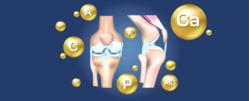
Calcium availability depends not only on intake but on absorption and distribution, and nutritional synergy plays a decisive role in this balance. The body relies on vitamin D to absorb calcium, on vitamin K to anchor calcium into skeletal structure, and on magnesium to stabilize calcium metabolism. Without these partners, calcium can be insufficiently absorbed or deposited in the wrong locations. A visual that shows calcium properly binding within bone alongside vitamin D, vitamin K, and magnesium icons helps reinforce that supplements or diets must support the full nutrient network, not calcium alone. Hydration, protein intake, and physical activity also determine how effectively calcium is converted into bone mass. Weight-bearing exercise stimulates mineral deposition, strengthening the skeleton where it encounters pressure. A vector illustration overlayed with movement imagery helps demonstrate this relationship—that muscles and bones are complementary systems, and physical activity directly reinforces mineral accumulation.
In holistic skeletal health education, the pairing of bone and marrow imagery with calcium flow creates a unified visual narrative: bones do not just house calcium; calcium enables bones to support biological life. Healthy bone structure promotes marrow health, and healthy marrow enables the body to function. When a person sees compact bone structure on one side, active marrow production on the other, and the pathway of calcium feeding both structural and metabolic needs, the purpose of skeletal nutrition becomes immediately understandable. The illustration simplifies a complex network into a coherent story of strength, density, and growth.
Ultimately, a bone and bone marrow vector illustration showing the importance of calcium becomes more than a diagram—it becomes a full explanation of how the skeleton develops, adapts, protects, produces, and sustains human life. By visualizing calcium not as an isolated mineral but as a continuous participant in structural and physiological systems, the illustration allows people of all backgrounds—students, patients, researchers, athletes, and clinicians—to understand why lifelong calcium intake, hormonal support, weight-bearing movement, and balanced nutrition safeguard mobility and independence. It highlights that strong bones do not happen by chance; they are built through knowledge, nourishment, and the daily care we give to the living organs responsible for strength and vitality throughout the body.