Iris Pigmentation Types Vector Illustration for Medical and Anatomy Education
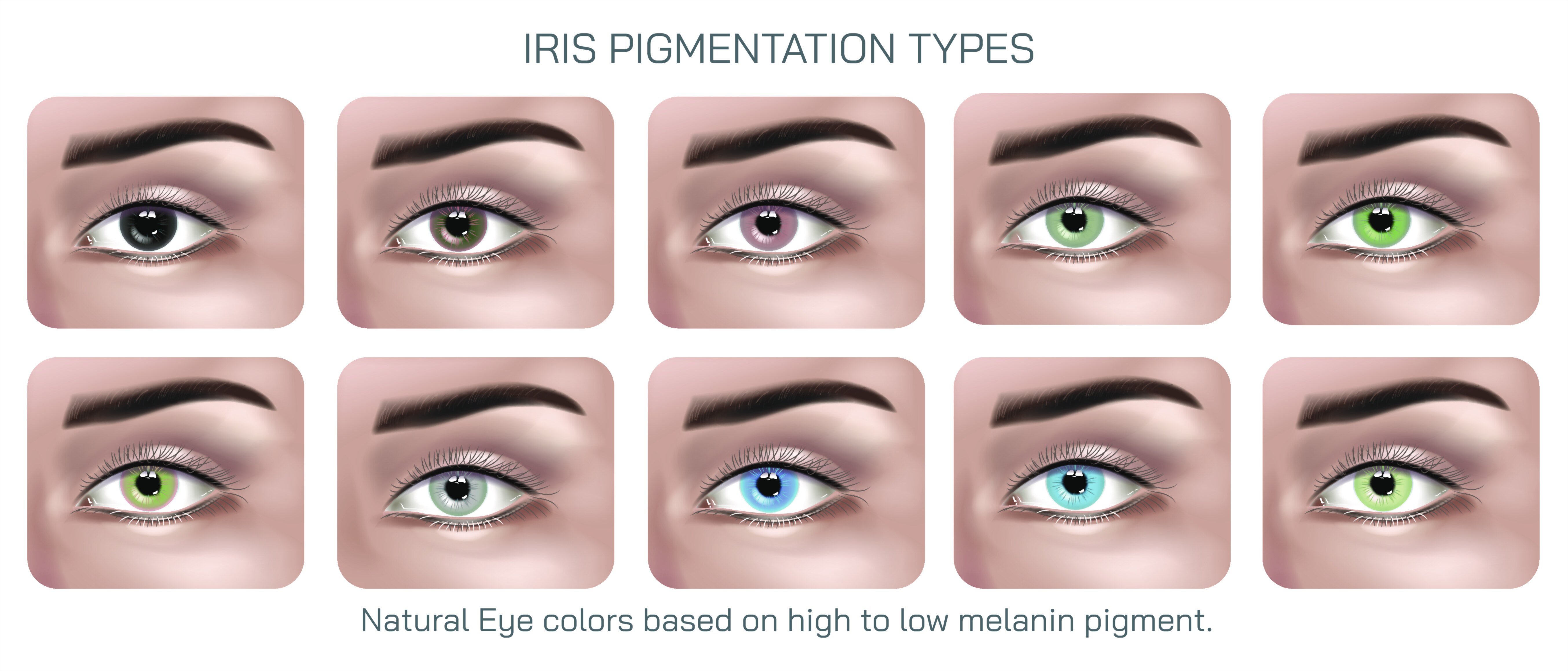
The human iris has long fascinated scientists, doctors, anatomists, geneticists, and educators because it is one of the most visually distinctive and biologically complex structures in the body. The wide variety of iris colors and patterns seen across the human population is not just a matter of aesthetics; it reflects underlying genetic mechanisms, melanin distribution, developmental biology, structural anatomy, and evolutionary history. When an iris pigmentation types vector illustration is designed for medical and anatomy education, it carries an important scientific purpose: to visually decode the subtle variations of pigmentation while maintaining anatomical accuracy and educational clarity. Such an illustration becomes a learning gateway for subjects ranging from genetics and ophthalmology to anthropology and medical imaging, enabling students and professionals to recognize how pigmentation differences correlate with cellular structures and the light-scattering properties within the iris stroma and epithelium. The vector graphic format, with its clean lines and sharp scalability, makes this content ideal for textbooks, e-learning modules, research presentations, medical infographics, academic posters, and anatomy lectures where precision and clarity shape successful understanding.
The iris is far more than a colorful surface; anatomically, it acts as a dynamic diaphragm responsible for controlling the size of the pupil. It enables smooth transitions between bright and dim light by regulating the amount of light entering the eye. Beneath this physiological function lies a layered architecture that directly influences its pigmentation. The anterior border layer, the iris stroma, and the posterior pigment epithelium interact with melanin concentration and collagen fibers to yield the wide spectrum of colors observable in the population. A scientifically accurate illustration of iris pigmentation types must reflect these structural realities—not simply showing hues, but representing how density and distribution of melanosomes contribute to each color category. For example, a light blue iris does not contain blue pigment; instead, it results from low melanin presence and the scattering of light within the stromal fibers, a phenomenon that creates the optical illusion of blue. Meanwhile, brown irises contain high concentration of melanin distributed throughout the stroma and pigment epithelium, resulting in deeper, darker coloration.
Medical and anatomy-focused vector illustrations of iris pigmentation therefore serve an essential educational role by visually categorizing primary pigmentation types. Blue irises symbolize low stromal melanin, green typically indicates intermediate levels of melanin blended with light-scattering effects, hazel represents a gradient of brown and amber tones with uneven melanin distribution, and gray reflects light scattering combined with altered collagen density. Amber has its own distinct identity, created not by low or high melanin overall but by a predominance of pheomelanin that produces warm golden or copper hues. Dark brown and near-black irises reflect the densest melanin concentration and often appear most prevalent in global populations, while very pale blue, gray, or depigmented iris patterns may correspond to genetic variations, congenital conditions, or albinism. When these variations are illustrated clearly, students of medicine and anatomy can connect visual differences with underlying biological mechanisms, making the iris not just an observational trait but a scientific marker.
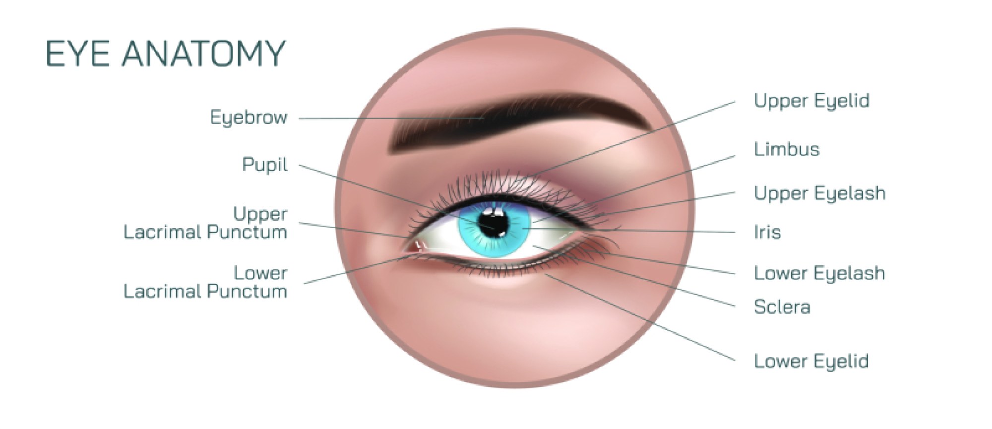
Vector illustrations of iris pigmentation types are particularly valuable because they deliver anatomical features without distortion, pixelation, or inaccuracies that commonly occur in photographs. Medical educators benefit from being able to enlarge or reduce the illustration on different screen sizes or printed surfaces without losing detail, allowing the position of crypts, radial furrows, contraction wrinkles, pupillary ruffs, and stellate patterns to remain visible. These morphological elements of the iris are often more striking in lighter pigmented eyes where structural contrast becomes noticeable, but they exist in all pigmentation types and therefore should be represented with equally careful attention. Even differences in iris texture — whether smooth, reticulated, velvety, or fiber-rich — can be translated into vector strokes and gradients, giving learners insight into subtle functional and diagnostic cues used in ophthalmologic examination.
Beyond anatomical accuracy, iris pigmentation illustrations serve an important role in teaching genetics. Students studying inheritance patterns are frequently taught that iris color is polygenic — influenced not by a single gene but by multiple gene interactions affecting melanin transport and synthesis. Modern genetics has moved beyond oversimplified dominant-recessive models to recognize the complex contributions of genes such as OCA2, HERC2, TYR, SLC24A4, and others in determining pigmentation levels. Having a visual reference of different pigmentation types helps learners internalize the concept that eye color is not binary, but exists on a rich spectrum shaped by population history and adaptive evolution. For example, lighter pigmentation is observed more frequently in populations historically from northern climates with lower UV exposure, whereas darker pigmentation predominates in equatorial populations where melanin offers protective benefits against ultraviolet damage. These relationships allow iris illustration to support deeper discussions of human migration, adaptation, genetic drift, and environmental pressures.
Another dimension that makes iris pigmentation educationally valuable is its clinical relevance. Eye color and pigmentation intensity can play a role in susceptibility to certain ocular conditions. Individuals with lighter iris pigmentation may be more prone to photophobia, macular degeneration risks linked to UV sensitivity, or pigment dispersion issues, while darker pigmentation can increase risk of pigmentary glaucoma or affect the appearance of clinically significant vascular changes. A detailed vector illustration showing pigmentation variations enables medical students to visually associate iris color with differential risk factors and diagnostic patterns. It becomes a preclinical teaching reference that builds visual memory before hands-on clinical experience.
When used in digital medical course design, iris pigmentation vector illustrations also bridge science and design in a compelling way. They help educators present anatomy in a manner that is both scientifically rigorous and visually engaging, encouraging learners to observe rather than memorize. By highlighting structural realism without overwhelming visual noise, a vector illustration supports memorization through familiarity rather than rote learning. It can also integrate seamlessly into interactive environments — such as clickable modules that allow students to explore pigmentation variations layer by layer, compare iris types side-by-side, or examine pigmentation under different lighting simulations. These dynamic uses transform the illustration into an instrument of discovery and critical thinking.
The emotional and psychological dimension of iris pigmentation adds another unique educational layer. Although teaching materials must always remain medically neutral and fact-based, students often connect personally when studying eye anatomy because the iris represents individuality and identity. No two irises are exactly alike — not even in identical twins — making the iris a biological signature as distinctive as a fingerprint. When learners view vector illustrations of diverse iris types, it reinforces the understanding that anatomy exists not only in theory but in real human variation. This cultivates respect and scientific curiosity about human diversity, which is valuable for both medical and research ethics.
In an academic context where clarity, accuracy, and engagement are critical, the iris pigmentation types vector illustration becomes far more than a graphical asset. It becomes a learning tool that integrates anatomy, physiology, genetics, ophthalmology, anthropology, and clinical observation. It supports classroom lectures, digital courses, medical discussions, and visual diagnostics by enabling students and practitioners to recognize how small differences in pigmentation patterns correspond to biological mechanisms with meaningful implications. As medical and anatomy education continues to embrace multimedia-assisted learning, this type of illustration stands as a vital resource — not only teaching what the iris looks like, but revealing how and why it varies from one person to another, and what that means for human health, identity, and evolutionary history.