Strabismus Surgery Vector Illustration for Eye Disease
Strabismus surgery is a highly specialized ophthalmologic procedure designed to correct eye misalignment by adjusting the strength, tension, or positioning of the eye muscles that control ocular movement. For medical and education purposes, a vector illustration of strabismus surgery serves as an invaluable visual tool because it transforms an internal biomechanical process into a clear, accessible, and precise learning model. Strabismus, often referred to as “crossed eyes” or “wandering eye,” occurs when the eyes do not align properly and therefore fail to coordinate their movements. This lack of alignment disrupts binocular vision and depth perception, and if left untreated during childhood, it can lead to amblyopia and long-term vision impairment. A surgical vector illustration provides an organized visual explanation of how surgery restores alignment, what structures are being modified, and how muscle balance is recalibrated to support clearer, coordinated vision.
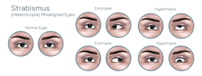
A typical educational illustration begins by showing the anatomy of the extraocular muscles—the six muscles that control horizontal, vertical, and rotational movement of the eye. In strabismus, one or more of these muscles may exert too much or too little pull, causing one eye to deviate inward (esotropia), outward (exotropia), upward (hypertropia), or downward (hypotropia). A vector graphic can clearly label the medial rectus, lateral rectus, superior and inferior rectus, and the superior and inferior oblique muscles. Highlighting the imbalanced or overactive muscle visually helps trainees and patients understand why the eye turns in a certain direction. Because vector illustrations retain perfect clarity at any scale, they are ideal for showing surgical steps that require millimeter-level precision.
Strabismus surgery typically involves one or more of three main techniques: recession, resection, and plication. A recession procedure weakens an overacting muscle by detaching it from its original insertion and reattaching it farther back on the sclera, thereby reducing its pulling power. A resection procedure strengthens a weak muscle by removing a small section of muscle tissue and reattaching the shortened muscle closer to the front of the eye, increasing its pulling force. Plication is an alternative to resection in which the muscle is folded and sutured to increase tension without cutting tissue. A vector illustration can depict each of these surgical concepts step-by-step: scalpel placement, disinsertion, suture positioning, repositioning of the muscle, and final tightening. This visual breakdown is particularly useful in surgical training, helping residents understand how altering muscle geometry redistributes the forces that align the eyes.
Some strabismus surgeries involve adjustable sutures, allowing the surgeon to fine-tune muscle tension after the patient awakens from anesthesia. A vector model can visually demonstrate adjustable knot loops, sliding sutures, and post-operative adjustment, which is much harder to conceptualize through text alone. For complex strabismus cases—such as paralytic strabismus, restrictive thyroid eye disease, or re-operation after previous muscle surgery—vector diagrams can show tethering, fibrosis, or altered muscle paths to guide advanced decision-making.
Another crucial benefit of vector illustrations for strabismus surgery is the ability to highlight how the procedure restores binocular visual function rather than merely cosmetic appearance. When misalignment is corrected, the visual axes of both eyes converge at the same target, allowing the brain to fuse the images into a single perception. An educational vector diagram can include before-and-after comparisons—showing double vision pathways before surgery and unified image fusion afterward—to reinforce the neurological impact of correction. Simulated before-surgery images can demonstrate how strabismus causes blurred, doubled, or overlapping fields of view, while post-surgery visuals can show improved alignment, depth perception, and visual comfort.
Beyond anatomy and surgery mechanics, vector illustrations can educate families on post-operative recovery and expectations. Many parents worry when their child wakes up with redness, subconjunctival swelling, or temporary over- or under-correction. An educational diagram that explains these phenomena visually—showing how the eye heals gradually and how alignment can continue to improve as muscles adapt—helps reduce anxiety and supports realistic expectations. For patients who require amblyopia therapy after surgery, illustrations can also show how improved alignment facilitates visual rehabilitation by restoring balanced sensory input to the brain.
In surgical education, vector graphics offer several advantages over photographs and microscope images:
• They eliminate distracting reflections and lighting artifacts.
• They allow selective highlighting of key anatomical layers—muscle, sclera, conjunctiva—at the instructor’s discretion.
• They can be animated or layered to show progressive steps without losing detail.
Vectors also support communication with non-medical audiences. For example, simplified versions of the illustration can appear in clinic brochures, videos, or pediatric consult discussions, helping caregivers understand why surgery is recommended and how it is performed safely.
Ultimately, a strabismus surgery vector illustration is far more than a diagram. It functions as a comprehensive visual educational resource that brings together ocular muscle anatomy, biomechanics of eye movement, neurological principles of binocular vision, surgical methodology, and post-operative outcomes. By making the invisible mechanics of extraocular muscle imbalance visible, the illustration deepens understanding in surgical trainees, fosters informed decision-making for patients and families, and contributes to more effective communication across the entire field of ophthalmology.