Types and Angles of Injections — Medical Administration Guide
The administration of injections is one of the most fundamental and widely practiced procedures in clinical medicine, yet it represents a complex interplay of anatomy, pharmacology, physiology, and technique. When examining the types and angles of injections used in medical settings, it becomes clear that every method is shaped by deep principles that govern how medications enter the body, how tissues respond to puncture and absorption, and how therapeutic agents must be placed to achieve both safety and therapeutic efficacy. Injections bypass many of the limitations of oral drugs, offering rapid delivery, precise dosing, and direct access to specific body compartments. The choice of injection route depends on the nature of the drug, the urgency of treatment, patient-specific factors, and the anatomical structure of the targeted tissue. The angle of needle insertion further determines how deeply the medication is deposited, which tissue layer it enters, and how effectively it will be absorbed. Understanding the conceptual foundations of these injection types reveals how carefully orchestrated medical procedures must be to maintain safety, minimize discomfort, and ensure predictable clinical outcomes.
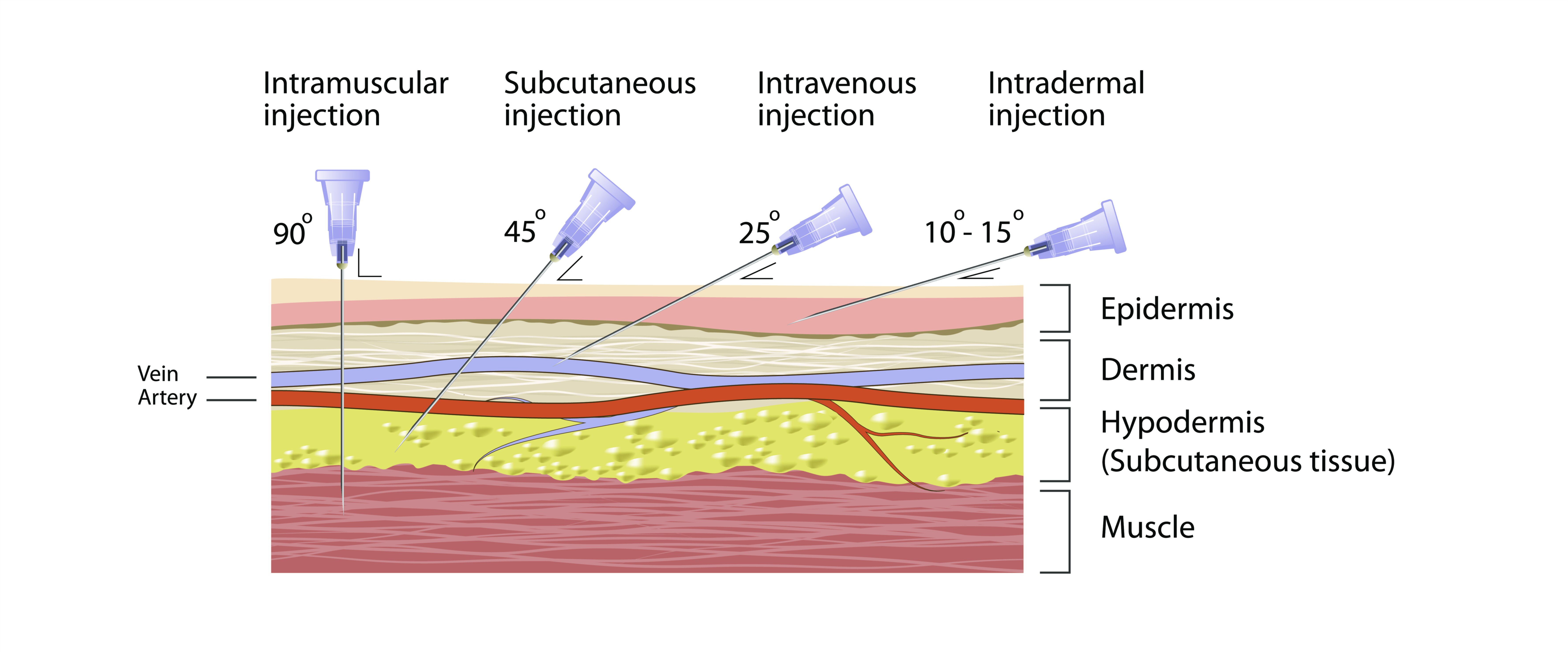
Among the most common injection types is the intramuscular injection, a method that places medication into the muscle tissue beneath the subcutaneous layer. Muscles are well-vascularized tissues, allowing medications to enter systemic circulation more rapidly than they would through subcutaneous fat. This makes intramuscular injections ideal for vaccines, hormones, antibiotics, analgesics, and medications that require intermediate absorption rates. The angle of needle insertion is typically steep, entering at around ninety degrees relative to the skin. This perpendicular approach ensures that the needle penetrates the epidermis, dermis, and subcutaneous fat before entering the muscle fibers themselves. The structure of muscle tissue—with its abundant capillary networks and thick bundles of contractile fibers—supports steady and efficient absorption. Because intramuscular injections must reach deeper layers, clinicians select specific anatomical sites such as the deltoid, vastus lateralis, or ventrogluteal muscle. These areas provide sufficient muscle mass while minimizing the risk of injuring major nerves or blood vessels. The choice of site reflects a deep anatomical knowledge, ensuring that each injection balances safety with therapeutic efficiency.
Subcutaneous injections represent a second major category, involving the delivery of medication into the fatty tissue just beneath the skin. Subcutaneous fat contains fewer blood vessels than muscle, which slows absorption and allows medications to diffuse more gradually into the bloodstream. This slower uptake is desirable for drugs that benefit from sustained release, such as insulin, certain anticoagulants, and various hormones. The angle of insertion for subcutaneous injections varies depending on the thickness of the patient’s adipose layer, typically falling between forty-five and ninety degrees. Thinner patients may require a more shallow approach to avoid penetrating too deeply, while individuals with thicker subcutaneous layers can safely receive injections at steeper angles. The flexibility of technique reflects the adaptability required when treating diverse patient populations. Common sites include the abdomen, upper arms, thighs, and lower back, regions selected for their accessibility and relative consistency in fat distribution. The subcutaneous environment offers a stable medium for drugs designed for prolonged absorption, illustrating how physiology determines technique.
Intravenous injections form another essential method, delivering medication directly into the bloodstream through a vein. This route bypasses tissue absorption entirely, offering immediate therapeutic effects and precise control over dosing. Because venous structures lie close to the surface, the angle of insertion is typically shallow, often around twenty-five degrees or less. This shallow trajectory helps guide the needle smoothly into the vein without penetrating through the vessel wall. Intravenous injections and catheter placements require careful visualization or palpation of veins, knowledge of venous patterns, and attention to patient factors such as hydration, vascular fragility, and age. The rapid onset of drug action through this route makes it indispensable in emergency medicine, critical care, anesthesia, and many diagnostic procedures. Yet intravenous administration also carries risks—including infiltration, infection, phlebitis, and rapid drug reactions—illustrating that precise angles and proper technique are essential for patient safety.
Intradermal injections represent a finer and more delicate method than the previous types, placing medication just beneath the surface of the skin, within the dermal layer. The dermis contains dense networks of immune cells, making this route particularly useful for sensitivity tests, allergy screening, and certain vaccines where controlled exposure to antigens is necessary. Intradermal injections use an extremely shallow angle of insertion, often around ten to fifteen degrees, allowing the needle to slip parallel to the skin surface. As medication is deposited into the dermis, a small raised wheal or blister-like area typically forms, indicating proper placement. This technique requires gentle precision, as injecting too deeply would enter subcutaneous tissue and disrupt both the diagnostic accuracy and the intended immunological response. The forearm is a common site for these injections due to its accessibility and thin dermal layer, making the resulting wheal easier to observe and evaluate.
Intraosseous injections represent a specialized method used primarily in emergency or critical situations where intravenous access is difficult or impossible. This technique involves inserting a needle directly into the bone marrow cavity, allowing medications and fluids to enter the central circulation almost as rapidly as through a vein. The bone marrow is richly vascularized, acting as an internal reservoir of blood supply capable of supporting urgent therapeutic delivery. Because the target lies within the bone, the angle of insertion is not defined by degrees but rather by anatomical landmarks and the orientation of bone surfaces. The procedure requires specialized needles capable of penetrating cortical bone. Intraosseous lines are typically placed in the tibia, humerus, or sternum during life-saving interventions when shock, dehydration, or vascular collapse prevents access to veins. This route shows how medical technique adapts to the physiological challenges present in critical care.
Intrathecal injections deliver medication into the cerebrospinal fluid surrounding the spinal cord. This highly specialized procedure requires precise anatomical knowledge and is often performed to administer chemotherapy, anesthesia, or diagnostic agents. The angle of insertion depends on spinal positioning and must be guided carefully between vertebrae to avoid damaging nerves. Because the cerebrospinal fluid environment is highly sensitive, intrathecal injections require absolute sterility, steady technique, and an understanding of the fluid’s circulation patterns. Even slight deviations can have serious neurological consequences, demonstrating how profoundly injection angles and anatomical precision matter in advanced medical procedures.
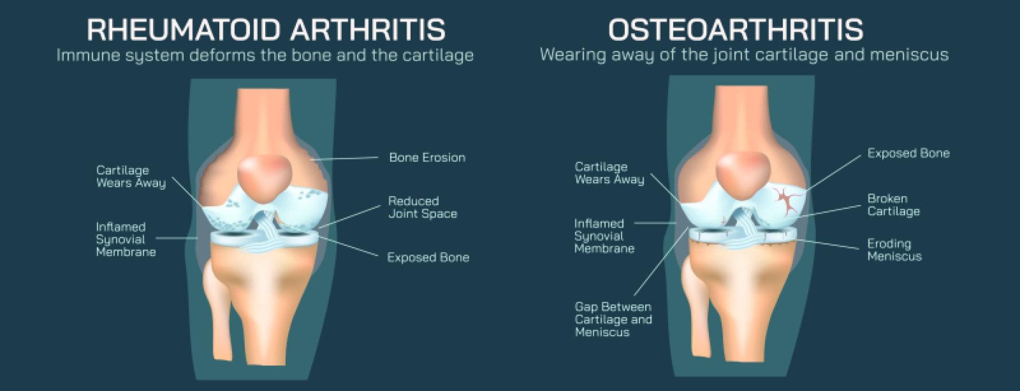
Intra-articular injections place medications directly into joint spaces, treating conditions such as arthritis, inflammation, or chronic joint pain. The joint cavity is surrounded by bone, cartilage, synovial membranes, and ligaments, meaning that the injection must be oriented based on the geometry of the joint itself. The angle of insertion varies depending on the joint—knees, shoulders, hips, and wrists each require different approaches to avoid damaging cartilage or other structures. The medication used, often corticosteroids or hyaluronic acid, must be delivered into the synovial fluid compartment where its therapeutic influence is concentrated. This route reflects the deep relationship between anatomical architecture and procedural technique.
Intraperitoneal injections introduce medication into the peritoneal cavity, the expansive space surrounding abdominal organs. While not commonly used in everyday clinical practice for humans, this method appears in certain chemotherapy treatments and is widely used in research environments with laboratory animals. The angle and site of insertion must avoid major organs, requiring anatomical awareness of abdominal wall thickness and organ positioning. This route provides access to a large absorptive surface, allowing medications to enter circulation gradually through peritoneal membranes.
Understanding the angles and types of injections also requires acknowledging physiological responses to puncture. The skin, fat, muscle, and vascular tissues each possess distinct densities, sensitivities, and biochemical properties. The angle at which a needle enters determines which of these layers are accessed and influences the patient's comfort, the likelihood of complications, and the efficacy of drug delivery. For example, incorrect angles during intramuscular injections may lead to shallow placement, sending the drug into subcutaneous fat instead of muscle, slowing absorption and reducing therapeutic effectiveness. Conversely, an overly steep or poorly positioned injection may strike nerves or blood vessels, causing pain or injury. Similarly, improper angles in intravenous injections may lead to extravasation, where medication leaks into surrounding tissue rather than entering the bloodstream. Each error highlights why precise technique rooted in anatomical and physiological understanding is indispensable.
The conceptual importance of injection types reaches far beyond the mechanics of needle insertion. These methods reflect the deeper biological truth that the human body is composed of layers with distinct functional properties. Medications must be strategically placed in the correct compartment to behave predictably. Through intramuscular injections, clinicians rely on vascular muscle tissue to deliver drugs efficiently. Through subcutaneous injections, they use slower-absorbing fat layers to achieve sustained therapeutic levels. Through intravenous and intraosseous routes, they leverage direct entry into the circulatory network for rapid systemic effects. Through intradermal and intra-articular injections, they harness localized environments for targeted diagnostics or treatments. In each case, the body’s structure shapes clinical decisions, and the needle angle becomes a technical expression of deeper biological and pharmacological principles.
Ultimately, the types and angles of injections are far more than procedural steps—they represent the merging of anatomical knowledge, clinical judgment, patient safety, and therapeutic strategy. Each injection type demonstrates how medicine transforms the abstract concepts of physiology into practical, life-supporting interventions. Every angle and technique reflects a thoughtful translation of biological structure into effective treatment. Understanding these injection methods deepens our appreciation not only for the science of drug delivery but for the precision, intentionality, and anatomical insight that underpin even the most routine medical actions.