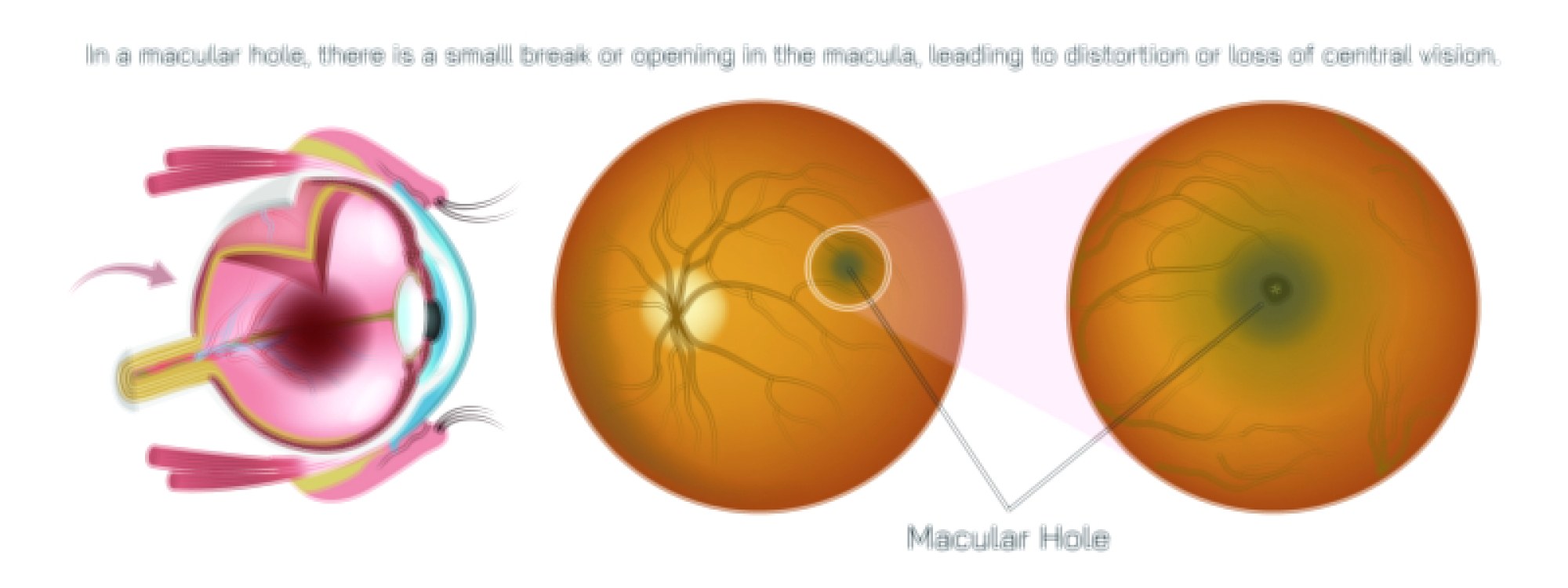
Human Chronic Kidney Disease Vector Illustration Showing Damaged Kidneys, Reduced Function, and Associated Health Complications
Chronic kidney disease (CKD) is a progressive condition characterized by gradual loss of kidney function over time, leading to the accumulation of waste products, electrolyte imbalances, and fluid dysregulation in the body. Understanding CKD’s anatomy, pathology, and systemic effects is crucial for medical education, nephrology, and public health awareness. A vector illustration depicting human chronic kidney disease typically integrates damaged kidneys, reduced renal function, affected nephrons, and associated health complications, providing a comprehensive and visually intuitive educational tool. By combining labeled kidney structures, functional impairment markers, and systemic effects, such diagrams help learners grasp both local renal pathology and its broader physiological consequences.
At the center of the illustration are bilateral kidneys, depicted in an anterior or lateral view with surrounding anatomical landmarks for context, including the renal arteries, veins, ureters, and adjacent organs. The kidneys are illustrated with a color gradient showing healthy tissue in normal shades of red and brown, contrasted with damaged regions highlighted in darker or mottled tones, representing fibrosis, scarring, or nephron loss. Labels identify renal structures such as the cortex, medulla, renal pelvis, glomeruli, and nephrons, linking structural anatomy to functional impairment. Arrows may indicate reduced blood flow or impaired filtration, visually emphasizing the impact of CKD on kidney physiology.
A critical aspect of the illustration is the representation of nephron damage. Vector insets magnify individual nephrons, showing glomerular sclerosis, tubular atrophy, and interstitial fibrosis. Labels indicate loss of glomerular filtration capacity, reduced tubular reabsorption, and impaired waste excretion. Arrows may illustrate reduced filtration rate, highlighting the pathophysiological basis of CKD symptoms such as uremia, electrolyte imbalance, and fluid retention. Color coding distinguishes healthy nephrons from damaged or nonfunctional units, providing a clear visual comparison.
The illustration often includes markers of systemic complications associated with CKD. For example, arrows and labels may point to fluid accumulation in the lungs and extremities, hypertension, anemia due to reduced erythropoietin production, and bone mineral imbalance resulting from impaired vitamin D metabolism. Color-coded icons or overlays may indicate affected organs such as the heart (cardiovascular complications), skeletal system (renal osteodystrophy), and blood vessels (vascular calcification), demonstrating how kidney dysfunction affects overall health.
Functional impairment is illustrated using flow indicators, such as arrows showing slowed glomerular filtration, reduced urine output, and toxin accumulation in the blood. Vector diagrams may also depict proteinuria by showing excess protein in urine, along with labeled pathways of waste buildup like creatinine and urea retention. These visual elements link anatomical kidney damage to measurable laboratory findings and clinical symptoms, enhancing educational relevance.
The illustration often includes progressive stages of CKD, from mild kidney damage (slightly reduced glomerular filtration rate) to severe or end-stage kidney disease requiring dialysis or transplantation. Side-by-side panels may show normal kidneys, early-stage CKD, and advanced CKD with shrunken, scarred tissue. Arrows between stages indicate disease progression, emphasizing the chronic nature of kidney dysfunction and the importance of early detection and intervention.
Additional educational features may include renal blood supply and filtration dynamics. Arrows illustrate impaired perfusion from narrowed or damaged renal arteries, while magnified cross-sections show glomerular capillaries affected by sclerosis or hypertension. Labels indicate how these changes reduce filtration efficiency and disrupt electrolyte and fluid balance. Color-coded overlays may differentiate oxygenated blood from poorly perfused areas, linking structural damage to functional consequences.
By combining bilateral kidney depiction, nephron-level damage, reduced filtration flow, systemic complications, disease progression stages, and functional markers, a vector illustration of human chronic kidney disease provides a comprehensive understanding of renal pathology. Color coding, labeled structures, magnified insets, and directional arrows allow learners to visualize how local kidney damage translates into systemic health effects, bridging anatomy, physiology, and pathology.
Ultimately, a CKD vector illustration demonstrates the connection between kidney structural damage, functional impairment, and systemic complications, transforming complex medical information into an educational and visually engaging tool. Through labeled kidneys, nephron details, impaired filtration indicators, and systemic health markers, the diagram makes chronic kidney disease comprehensible for medical students, healthcare professionals, and public health education.