The human skull anatomy represents one of the most complex and vital structural components of the skeletal system. The skull forms the bony framework of the head and performs several essential functions, including protecting the brain, supporting facial structures, housing sensory organs, and enabling functions such as chewing, speaking, and breathing. A detailed understanding of the skull’s structure is fundamental in biology, anatomy, medicine, dentistry, and forensic science.
The human skull is composed of multiple bones that are tightly joined together, forming a strong yet lightweight protective structure. In adults, the skull consists of twenty-two major bones that are broadly classified into two groups: cranial bones and facial bones. These bones are connected primarily by immovable joints known as sutures, which provide strength while allowing limited growth during early life.
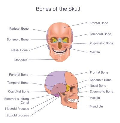
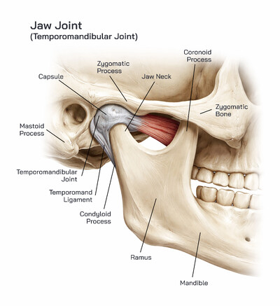
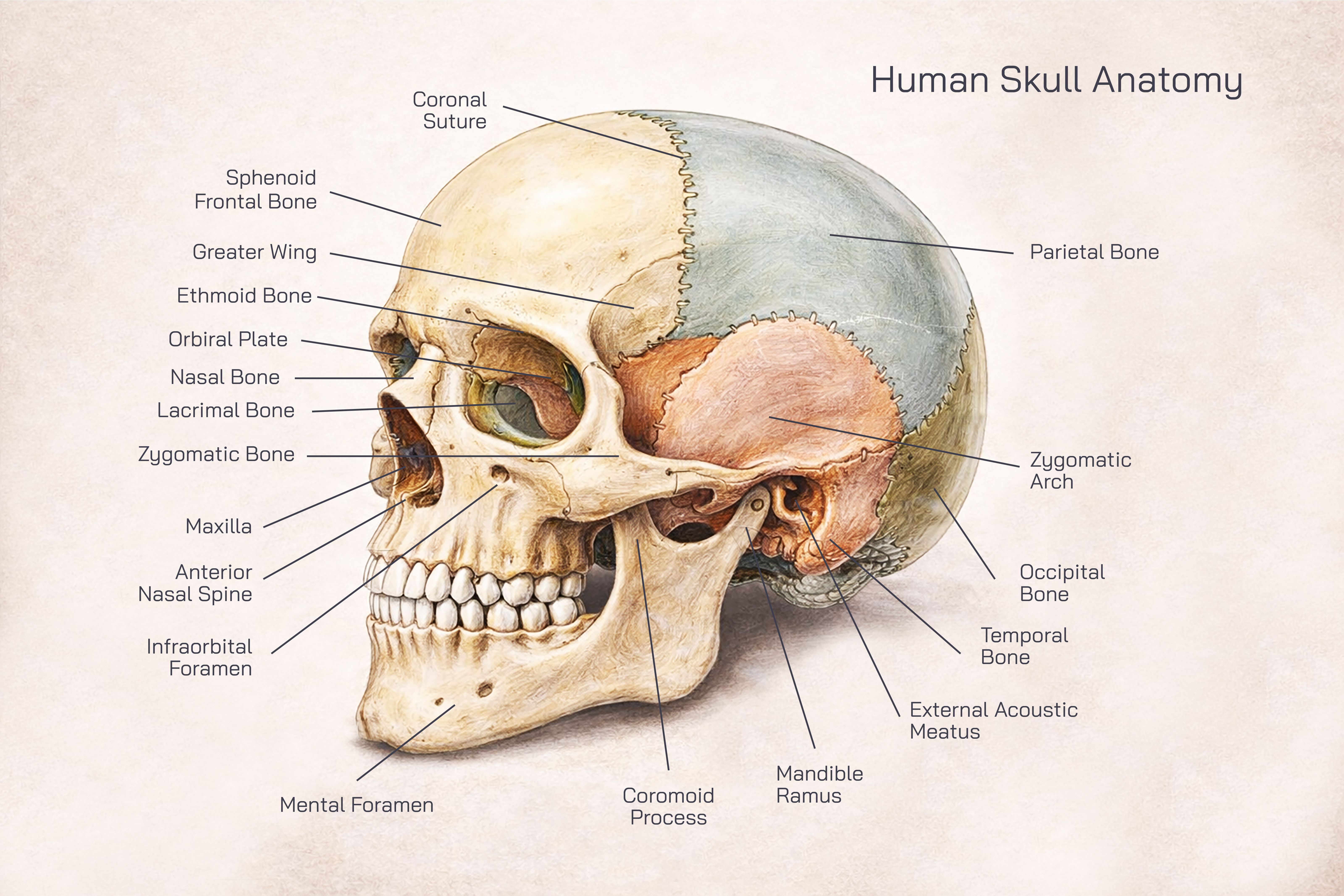
The cranial bones form the cranium, which encloses and protects the brain. There are eight cranial bones in total. The frontal bone forms the forehead and the upper part of the eye sockets. It also contributes to the roof of the nasal cavity. The parietal bones are two large bones that form the sides and roof of the cranium, playing a major role in enclosing the brain. The temporal bones are located on the sides of the skull and house important structures related to hearing and balance. They also form part of the jaw joint.
The occipital bone is located at the back and base of the skull. It contains a large opening through which the spinal cord passes to connect with the brain. This bone provides structural support and helps stabilize the head on the vertebral column. The sphenoid bone is a centrally located bone that helps connect the cranial bones together. Its unique shape allows it to support the brain and form part of the eye sockets. The ethmoid bone is a light, spongy bone located between the eye sockets. It contributes to the formation of the nasal cavity and eye orbits and plays a role in separating the nasal passages from the brain.
Facial bones form the framework of the face and support the features involved in eating, breathing, and facial expression. There are fourteen facial bones. The mandible, or lower jaw, is the only movable bone of the skull and is essential for chewing and speaking. The maxilla forms the upper jaw and holds the upper teeth. It also contributes to the formation of the nose, eye sockets, and hard palate.
The zygomatic bones, commonly known as cheekbones, give shape to the face and form part of the eye sockets. The nasal bones form the bridge of the nose, providing structure and support. The lacrimal bones are small and located near the inner corners of the eyes, helping form the tear ducts. The palatine bones contribute to the hard palate, separating the nasal cavity from the oral cavity. The inferior nasal conchae are curved bones inside the nasal cavity that help regulate airflow and humidify inhaled air. The vomer forms part of the nasal septum, dividing the nasal cavity into two chambers.
Sutures are an important feature of skull anatomy. These fibrous joints tightly bind skull bones together. Major sutures include the coronal suture between the frontal and parietal bones, the sagittal suture between the two parietal bones, the lambdoid suture between the parietal and occipital bones, and the squamous suture between the parietal and temporal bones. In infants and young children, sutures are flexible and allow skull expansion as the brain grows. Over time, these sutures gradually fuse to form a rigid protective case.
The skull also contains several cavities and openings that serve specific purposes. The cranial cavity houses the brain, while the orbital cavities protect the eyes. The nasal cavity supports breathing and smell, and the oral cavity contains the teeth and tongue. Numerous small openings allow nerves and blood vessels to pass between the brain and the rest of the body.
Functionally, the skull plays a critical role in protecting delicate structures. The thick cranial bones shield the brain from mechanical injury, while the facial bones protect sensory organs such as the eyes, nose, and mouth. The skull also provides attachment points for muscles responsible for facial expression, chewing, and head movement.
From a developmental perspective, skull anatomy reflects both protection and adaptability. At birth, skull bones are not fully fused, allowing the head to pass through the birth canal and accommodate rapid brain growth. As development progresses, the bones gradually fuse, providing increased strength and stability.
In medical and clinical contexts, knowledge of skull anatomy is essential. Doctors use skull landmarks for diagnosis, imaging interpretation, surgical planning, and trauma assessment. Dentists rely on detailed understanding of facial bones and jaw structure, while neurologists and neurosurgeons focus on cranial anatomy to protect brain function.
In conclusion, human skull anatomy consists of cranial and facial bones arranged in a precise and highly functional structure. Together, these bones protect the brain, support sensory organs, shape the face, and enable vital activities such as eating and speaking. The skull’s combination of strength, complexity, and adaptability makes it one of the most important components of the human skeletal system and a cornerstone topic in anatomy and medical education.