The human skull is one of the most remarkable structures of the skeleton. It provides a protective case for the brain, supports the senses of sight, smell, taste and hearing, and forms the framework of the face. Although it appears as a single solid object, the skull is actually composed of multiple bones that fuse together during development. Each bone has a unique shape, function and position, contributing to both protection and movement. Understanding the bones of the human skull is essential to understanding the structure of the head, the nervous system and the foundation of facial anatomy.
The skull is divided into two major groups of bones: cranial bones and facial bones. The cranial bones surround and protect the brain, forming the cranial cavity. The facial bones support the openings of the respiratory and digestive systems and shape the features of the face. Although the skull contains 22 bones in total, most of these bones are connected by immovable joints called sutures, which resemble cracks on the skull surface. These sutures stabilize the skull while allowing slight growth during childhood.
The cranial bones include eight bones arranged around the brain. The frontal bone forms the forehead and the roof of the eye sockets. Behind it lie the paired parietal bones, which make up the top and sides of the skull. On the lower sides of the head are the temporal bones, which house structures of the middle and inner ear essential for hearing and balance. At the back of the skull is the occipital bone, which encloses the foramen magnum— the large opening that allows the spinal cord to connect with the brain.
Deeper within the skull sit two more cranial bones. The sphenoid bone spans the width of the skull and supports the base of the brain, forming the central wedge-shaped foundation for the skull. The ethmoid bone lies between the eye sockets and contributes to the nasal cavity and the bony separation between the brain and the nose. These cranial bones collectively create a strong yet lightweight barrier that safeguards the central nervous system against trauma.
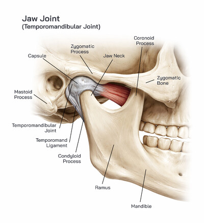
The facial bones give shape to the face, house the teeth and provide attachment points for the muscles used in expression, chewing and speaking. The mandible, or lower jaw, is the strongest bone of the skull and the only movable skull bone aside from the tiny bones of the middle ear. It supports the lower teeth and forms the chin. Above it lies the maxilla, the upper jawbone, which supports the upper teeth and forms part of the eye sockets and the palate.
In the midline of the face are the nasal bones, forming the bridge of the nose. The zygomatic bones, often called cheekbones, create the prominence of the cheeks and part of the outer orbit of the eyes. Behind the nose are the lacrimal bones, the smallest of the facial bones, containing a channel for tear drainage. The palatine bones form the back portion of the hard palate, separating the mouth from the nasal cavity. The slender vomer contributes to the nasal septum, dividing the nasal cavity vertically. The paired inferior nasal conchae curve inside the nasal passage, increasing the surface area for humidifying and filtering air.
The skull is held together by sutures, which appear as irregular wavy joints. The coronal suture separates the frontal bone from the parietal bones, the sagittal suture joins the two parietal bones along the midline, and the lambdoid suture connects the parietal bones to the occipital bone. The squamous suture lies between the parietal and temporal bones on each side. These sutures enable expansion during childhood and become nearly immovable in adulthood, providing long-term stability.
The skull does more than protect the brain — it contains openings and passageways that facilitate communication with the rest of the body. The foramina are small holes through which nerves and blood vessels travel. The orbits house the eyes and protect associated muscles, nerves and vessels. The nasal cavity is not only important for smell but also plays a role in filtering and warming air. The paranasal sinuses, present within the frontal, sphenoid, ethmoid and maxilla bones, reduce skull weight and contribute to voice resonance.
Structurally, the skull balances protection and functionality. Thick bony plates defend the brain, while lighter cancellous bone reduces weight. The jaw enables chewing and speech, while facial bones support sensory organs. This combination of defense, mobility and sensory support highlights how evolution has refined the skull into a compact and efficient design.
Injury or disease affecting the bones of the skull can impact multiple body systems. Fractures may threaten the brain, while malformations can affect breathing, speech or vision. Dental problems may originate from jaw misalignment or bone deterioration. Age-related changes such as bone thinning and tooth loss may alter the facial skeleton. Understanding skull anatomy helps clinicians diagnose trauma, neurological disorders, sinus conditions, dental problems and congenital conditions.
The bones of the skull are not merely a protective shell but a dynamic foundation for human identity and function. Their complex arrangement supports cognition, sensation, expression and survival. Each bone — whether large like the mandible or tiny like the lacrimal — plays a necessary role in supporting life.