The human placenta is one of the most extraordinary organs in biology because it forms temporarily during pregnancy and serves as the central connection between the mother and the developing fetus. It acts as the biological bridge that sustains new life by providing oxygen, nutrients and growth signals while simultaneously removing waste products. Even though it functions for less than one year, the placenta performs roles so complex and vital that life in the womb could not continue without it. Understanding its structure and function provides insight into the hidden world of pregnancy and the processes that enable fetal development long before birth.
Structurally, the placenta develops after implantation, when the embryo attaches to the uterine lining during early pregnancy. Once implanted, specialized cells from the embryo called trophoblasts invade the uterine wall and form the initial scaffolding of the placenta. Over time, the placenta expands and becomes anchored deeply into the uterus, ensuring a stable and secure connection. The placenta grows into a disc-shaped organ that eventually weighs about one-sixth as much as the newborn at birth. One surface of the placenta attaches to the uterine wall, while the other surface connects to the fetus through the umbilical cord.
The maternal side of the placenta consists of many subdivisions called cotyledons, each containing numerous blood-filled spaces called intervillous spaces. Maternal blood from the uterine arteries flows into these spaces, pooling around tree-like structures known as chorionic villi. These villi originate from the fetal side of the placenta and are covered with a layer of trophoblast cells that act as a selective barrier. Inside each villus are fetal blood vessels that branch from the umbilical cord. Importantly, maternal and fetal blood do not mix; instead, nutrients and gases must diffuse across the trophoblast membrane. This intricate exchange system supplies oxygen and nutrients to the fetus while allowing carbon dioxide and metabolic waste to be carried away.
In addition to nutrient and waste exchange, the placenta serves as an endocrine organ, producing hormones required to support pregnancy. One of the earliest hormones secreted by the placenta is human chorionic gonadotropin (hCG), which signals the body to maintain progesterone levels and prevent menstruation. The placenta also produces progesterone itself, which stabilizes the uterine lining and prevents premature contractions. Estrogen levels rise steadily due to placental production, stimulating maternal blood flow and preparing the body for childbirth and breastfeeding. Other hormones produced by the placenta help regulate fetal metabolism, maternal glucose balance and immune tolerance, demonstrating that the placenta acts not only as a transporter but also as a hormonal powerhouse.
The placental immune function is equally remarkable. The fetus carries genetic material from both parents, meaning the immune system of the mother might naturally treat it as foreign. The placenta prevents this from happening by acting as an immunological shield. The placental barrier allows protective maternal antibodies to cross into the fetus, boosting immune protection during late pregnancy and after birth, but prevents most harmful microbes from crossing. The placenta also expresses molecules that prevent the mother’s immune cells from attacking fetal tissues. This delicate immunological balance allows the fetus to remain protected while safely growing inside the maternal body.
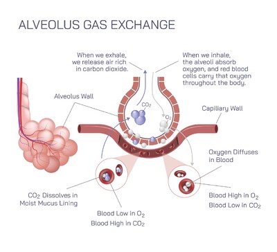
Placental circulation is the lifeline of pregnancy. Blood from the fetus travels through the umbilical arteries into the placenta, where carbon dioxide and waste diffuse outward into maternal blood. Oxygen and nutrients then diffuse inward from maternal blood into fetal circulation before purified fetal blood returns through the umbilical vein. The effectiveness of this exchange determines fetal growth and development, making healthy blood flow crucial. Conditions such as high blood pressure during pregnancy, placental insufficiency, or reduced uterine blood supply can impair nutrient and oxygen delivery, slowing fetal growth and increasing complications.
The placenta also adapts continually throughout pregnancy. Early in development, its primary role is to implant firmly and build a nutritional supply system. As the fetus grows, the number of chorionic villi increases, maximizing exchange surface area. Late in pregnancy, the placenta becomes more efficient at nutrient transfer to meet the high demands of rapid fetal growth. When childbirth approaches, the placenta begins preparing for delivery. After the baby is born, the placenta detaches naturally from the uterine wall and is expelled during the final stage of labor, marking the completion of its biological purpose.
Because the placenta influences every part of fetal development, its structural or functional abnormalities can have major consequences. Placental insufficiency, preeclampsia, placenta previa, placental abruption and infections can disrupt normal function and place both mother and baby at risk. Monitoring placental position, blood flow and hormone levels during pregnancy helps ensure that the organ continues to function properly. Healthy lifestyle choices — including good nutrition, regular prenatal care, avoidance of smoking and alcohol and management of maternal medical conditions — contribute to optimal placental function.
Despite functioning for a limited period, the human placenta is essential to life. It supports the fetus physically, metabolically, hormonally and immunologically, making pregnancy possible from implantation until birth. Through its sophisticated structure — from the chorionic villi and intervillous spaces to the umbilical cord and trophoblast barrier — the placenta achieves constant two-way communication between mother and fetus while maintaining strict biological separation. It is a temporary organ, but its role is enduring; the placenta lays the foundation for survival, growth and long-term health of the newborn.