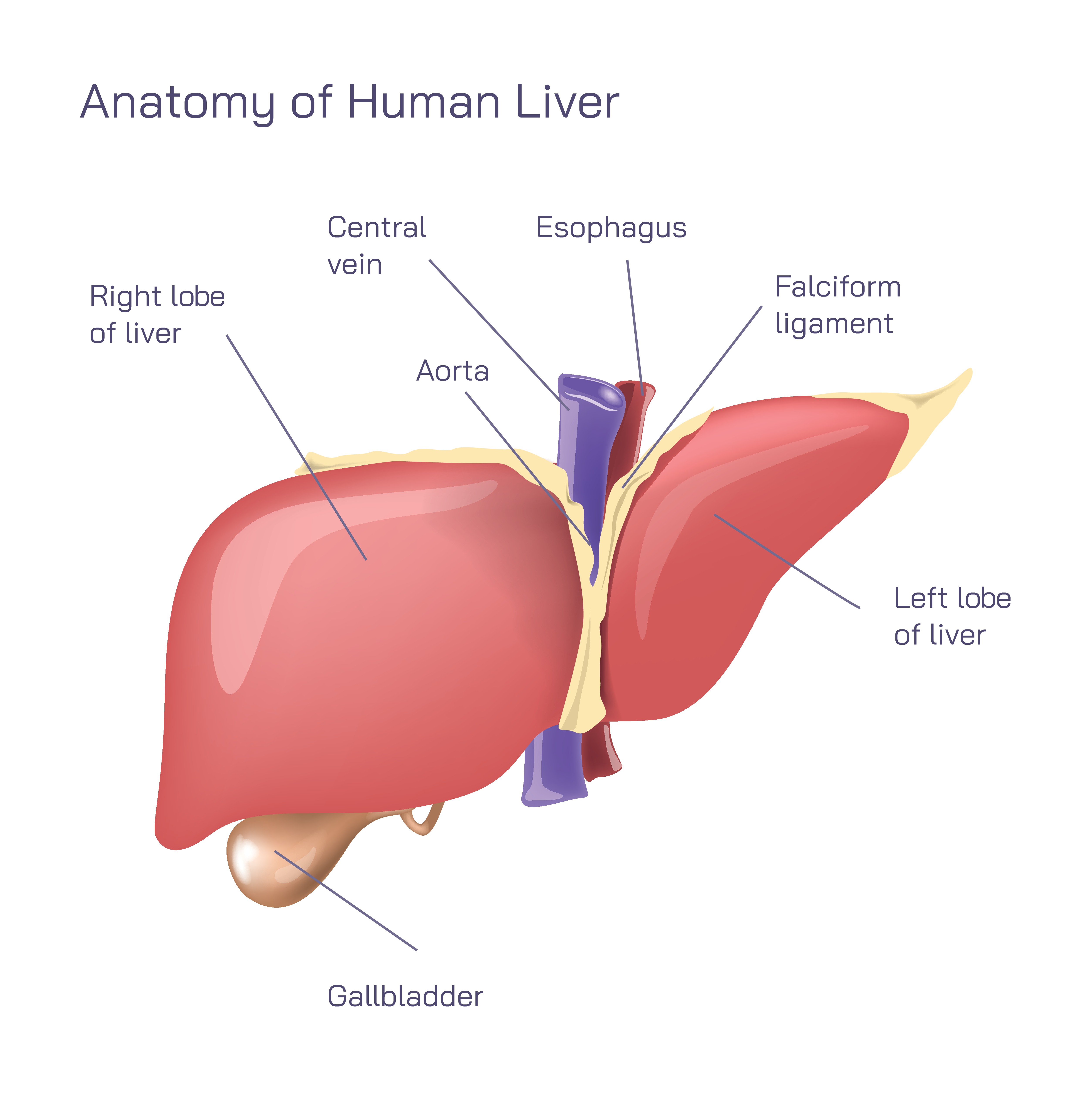
Anatomy of the Human Liver.
Understanding the Liver as a Vital Organ in Human Physiology
The liver is one of the most essential organs in the human body, performing hundreds of metabolic, detoxifying, and regulatory functions that sustain life. Located in the upper right portion of the abdominal cavity, just beneath the diaphragm, the liver plays a central role in digestion, nutrient processing, blood filtration, and chemical balance. Its unique structure enables it to handle large volumes of blood, process toxins, store nutrients, and produce bile, all while maintaining a regenerative capacity unmatched by most organs. Exploring the anatomy of the liver provides important insight into how its shape, lobes, vessels, and associated structures support its complex roles in the body.
Right and Left Lobes: Major Divisions of the Liver
The human liver is divided into two primary lobes: the right lobe and the left lobe. The right lobe is larger and occupies most of the upper abdominal area beneath the ribs, while the left lobe extends toward the left upper quadrant and is smaller in size. These lobes are separated by the falciform ligament, a thin fold of tissue that anchors the liver to the anterior abdominal wall. Although traditionally described as two lobes, functionally and surgically the liver is divided into eight segments based on its vascular supply. Regardless of interpretation, the right and left lobes provide a clear visual distinction and help clinicians identify regions for diagnosis or surgery.
The Falciform Ligament: A Structural Anchor
The falciform ligament is a sheet-like structure that attaches the liver to the diaphragm and the inside of the abdominal wall. It forms a natural dividing line between the right and left lobes. This ligament also contains the ligamentum teres, a remnant of the fetal umbilical vein that once carried oxygenated blood from the placenta. Although the falciform ligament does not perform metabolic functions, it plays an important anatomical role in positioning the liver securely within the abdominal cavity.
The Central Vein: A Key Vessel for Blood Drainage
Within the liver’s extensive network of blood vessels lies the central vein, which forms an essential part of the liver’s internal architecture. Blood from the hepatic sinusoids—tiny channels that filter blood—drains into the central veins, which merge into larger veins and ultimately flow into the hepatic veins. These hepatic veins empty into the inferior vena cava, returning cleansed and processed blood to the heart. This drainage system highlights the liver’s role in circulating nutrients and removing toxins from the bloodstream.
The Aorta and Esophagus: Important Neighboring Structures
Although the aorta and esophagus are not parts of the liver, they lie in close anatomical proximity. The esophagus runs behind the liver as it connects the throat to the stomach, and the aorta descends through the thorax to supply major abdominal organs, including the liver through the hepatic artery. These structures emphasize the density of organs and vessels in the upper abdomen and the functional interdependence between the liver and other parts of the digestive and circulatory systems.
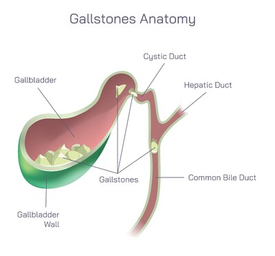
Gallbladder: Storage Site for Bile
Attached to the underside of the liver is the gallbladder, a small, pear-shaped organ that stores and concentrates bile produced by the liver. Bile is essential for digesting fats and absorbing fat-soluble vitamins. When food enters the small intestine, especially fatty foods, the gallbladder contracts and releases bile through the bile ducts. This close anatomical and functional connection between the liver and gallbladder forms an essential component of digestion and nutrient absorption.
The Liver’s Vascular Supply: Dual Blood Flow System
One of the most remarkable features of the liver is its dual blood supply. Blood enters the liver through two major vessels:
Hepatic Artery – Supplies oxygen-rich blood from the aorta.
Hepatic Portal Vein – Carries nutrient-rich blood from the digestive tract, spleen, and pancreas.
This dual input allows the liver to process nutrients absorbed from food, detoxify chemicals before they circulate throughout the body, and maintain stable blood composition. The filtered blood then leaves the liver through the hepatic veins.
Internal Microarchitecture and Functional Units
Each lobe of the liver is composed of thousands of microscopic units called lobules. These hexagonal structures contain hepatocytes—liver cells—that carry out most metabolic functions. The lobules organize blood flow in a radial pattern toward the central veins, ensuring efficient processing of hormones, drugs, nutrients, and waste materials. Their arrangement enables the liver to perform its work continuously and reliably.
A Multifunctional Organ with Critical Roles
The liver’s anatomy supports a wide range of physiological functions, including:
Detoxifying harmful substances
Producing bile for fat digestion
Storing minerals and vitamins
Regulating blood sugar levels
Synthesizing proteins such as albumin and clotting factors
Supporting immune defense
Breaking down old red blood cells
Its location, complex internal structure, and extensive vascular network make these functions possible.
The Liver’s Regenerative Ability: A Unique Biological Feature
Unlike most organs, the liver has a remarkable ability to regenerate after injury or surgical removal of a portion of its tissue. This regeneration allows it to maintain functionality even when damaged by disease, toxins, or surgery. Understanding its anatomy helps explain why the liver can adapt so effectively—its cellular organization and rich blood supply support rapid repair and regeneration.
An Essential Organ at the Center of Human Physiology
The human liver’s anatomy is a combination of structural elegance and functional complexity. Its lobes, vessels, ligaments, and associated organs work together to support digestion, metabolism, detoxification, and circulation. Recognizing the anatomy of the liver not only enhances knowledge of human biology but also provides insight into how vital and irreplaceable this organ is for maintaining overall health and well-being.