The human heart blood flow system is a highly organized and continuous process that ensures oxygen, nutrients, hormones, and waste products are efficiently transported throughout the body. The heart functions as a muscular pump that maintains circulation by rhythmically contracting and relaxing. A complete understanding of blood flow through the heart requires knowledge of its chambers, valves, and associated blood vessels, as well as the distinction between pulmonary and systemic circulation. The heart blood flow diagram visually represents this process and helps clarify how blood moves in a precise, unidirectional manner.
The heart is divided into four chambers: two upper chambers called atria and two lower chambers called ventricles. The right side of the heart is responsible for handling deoxygenated blood returning from the body, while the left side manages oxygenated blood that is delivered to tissues. This separation is essential for efficient circulation and is maintained by a muscular wall known as the interventricular septum.
Blood flow begins when deoxygenated blood from the body returns to the heart through large veins. The superior vena cava carries blood from the upper body, including the head, neck, and arms, while the inferior vena cava brings blood from the lower body. These veins empty into the right atrium. The right atrium serves as a receiving chamber and temporarily holds the blood before passing it to the right ventricle.
When the right atrium contracts, blood flows through the tricuspid valve into the right ventricle. This valve ensures that blood moves in only one direction and prevents backflow into the atrium. The right ventricle then contracts with sufficient force to push the deoxygenated blood out of the heart toward the lungs. As it contracts, blood passes through the pulmonary valve into the pulmonary trunk.
The pulmonary trunk divides into the right and left pulmonary arteries, which carry deoxygenated blood to the lungs. This stage marks the beginning of pulmonary circulation. In the lungs, blood flows through tiny capillaries surrounding the alveoli. Here, carbon dioxide is released from the blood, and oxygen is absorbed. This gas exchange process transforms deoxygenated blood into oxygen-rich blood, which is essential for cellular respiration.
Oxygenated blood returns to the heart through the pulmonary veins, which are unique among veins because they carry oxygen-rich blood. These veins deliver blood to the left atrium. The left atrium acts as a holding chamber and prepares the blood for transfer to the left ventricle, which is the strongest chamber of the heart.
When the left atrium contracts, blood passes through the mitral valve into the left ventricle. The mitral valve, also known as the bicuspid valve, ensures one-way flow and prevents blood from leaking backward into the atrium. The left ventricle has thick muscular walls because it must generate enough pressure to send blood throughout the entire body.
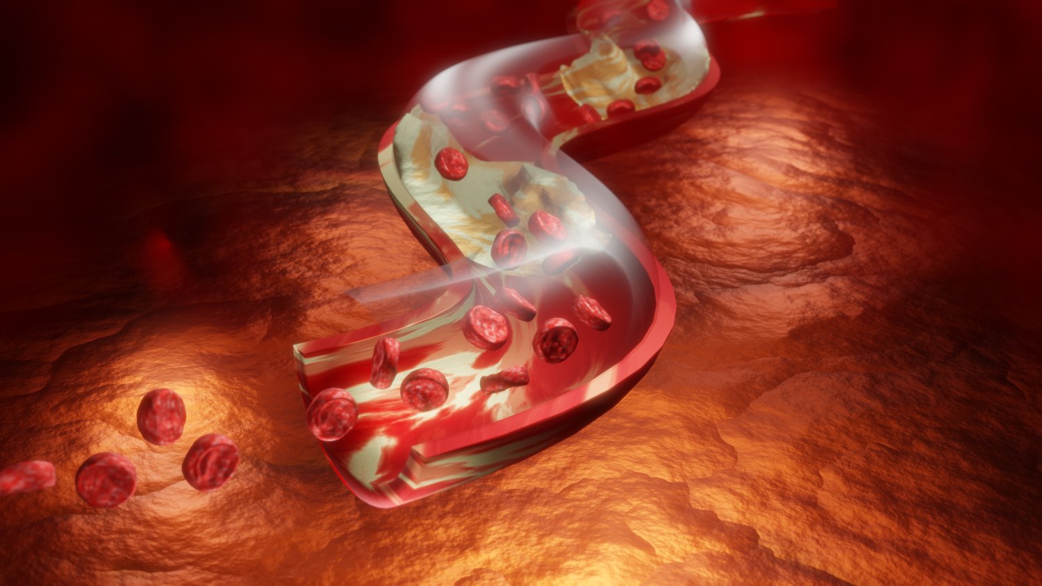
During ventricular contraction, oxygenated blood is forced through the aortic valve into the aorta, the largest artery in the body. This marks the beginning of systemic circulation. The aorta branches into smaller arteries that deliver oxygenated blood to various organs and tissues. From arteries, blood moves into arterioles and then into capillaries, where oxygen and nutrients are exchanged for carbon dioxide and metabolic waste.
After delivering oxygen and nutrients, blood becomes deoxygenated and begins its return journey to the heart. It flows from capillaries into venules and then into veins. These veins gradually merge to form larger veins, eventually returning blood to the heart through the superior and inferior vena cava. This completes one full cycle of blood circulation.
The heart valves play a crucial role in maintaining efficient blood flow. The tricuspid and mitral valves control blood movement between atria and ventricles, while the pulmonary and aortic valves regulate blood exiting the ventricles. These valves open and close in response to pressure changes within the heart chambers, ensuring smooth and unidirectional circulation.
The heart blood flow system operates continuously throughout life, adapting to the body’s needs. During physical activity, the heart beats faster and stronger to deliver more oxygen to muscles. During rest, the heart rate slows while still maintaining sufficient circulation. This adaptability highlights the efficiency and resilience of the cardiovascular system.
The separation of pulmonary and systemic circulation ensures that oxygenated and deoxygenated blood do not mix. This dual circulation system is a defining feature of the human heart and allows for high metabolic efficiency. The diagram of heart blood flow clearly demonstrates this separation and the coordinated movement of blood through each chamber and vessel.
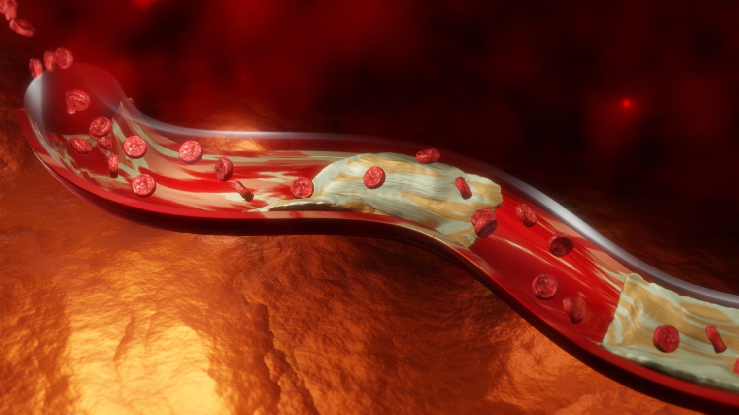
Understanding the human heart blood flow system is essential in biology, medicine, and health education. It provides insight into how the body maintains internal balance and supports all physiological functions. Disorders such as valve defects, blocked arteries, or weakened heart muscles disrupt this flow and can lead to serious health problems, emphasizing the importance of a well-functioning circulatory system.
In conclusion, the human heart blood flow system is a precise and continuous process involving four chambers, four major valves, and a network of blood vessels. Deoxygenated blood flows from the body to the right side of the heart, travels to the lungs for oxygenation, and returns to the left side of the heart before being pumped throughout the body. This coordinated circulation supports life by delivering oxygen and nutrients while removing waste products. The heart blood flow diagram serves as a clear and essential tool for understanding the structure, function, and importance of the cardiovascular system.