3D Rendered Neuron: A Detailed Visualization of Axons, Dendrites, and Synaptic Pathways
A 3D rendered neuron illustration offers one of the most immersive ways to understand the structure that drives thought, movement, sensation, and communication throughout the body. This digital representation captures the full complexity of a single neuron, revealing features that cannot be seen with the naked eye. By presenting these structures in a highly detailed visual format, the illustration helps viewers appreciate how this remarkable cell receives, processes, and transmits information with extraordinary precision.
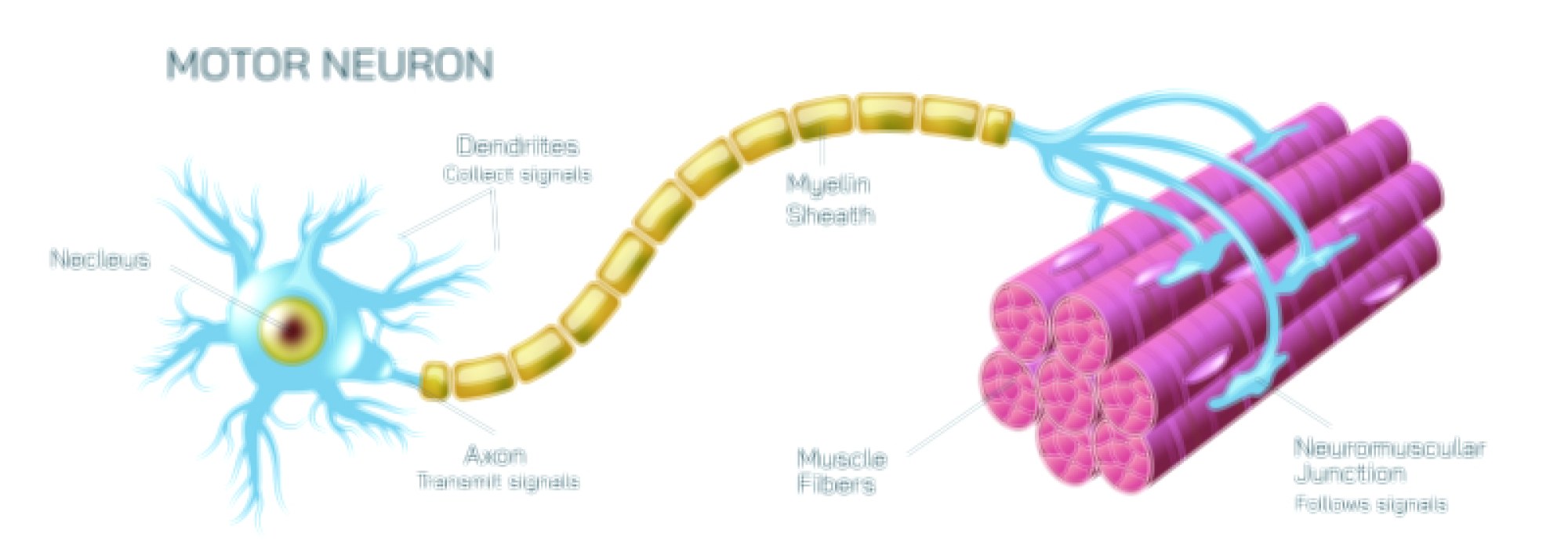
At the core of the illustration lies the cell body, the central hub where essential internal processes occur. The 3D rendering reveals the surface texture, gentle contours, and subtle lighting that symbolize the activity within. This region anchors the neuron’s communication system, acting as the site where important signals gather before being forwarded to distant regions. The detailed digital shading enhances the organic look, giving the viewer a sense of depth and physical presence.
From this central body extend the dendrites, beautifully branching structures that spread outward in numerous directions. In the illustration, these branches appear almost tree-like, capturing the intricate patterns that allow the neuron to receive information from many surrounding cells. The 3D format is especially effective here because it shows how each branch twists, angles, and stretches across space, emphasizing the cell's ability to participate in complex networks. The dendrites appear alive with detail, reinforcing the idea that they serve as the primary input channels for incoming signals.
Extending in the opposite direction is the axon—long, slender, and essential for output transmission. In the 3D depiction, the axon appears as a smooth, elongated pathway leading away from the cell body. Its curvature, surface glow, and layered texturing communicate its role as a high-speed route for outgoing messages. The illustration often includes subtle highlights along the axon to give a sense of direction and flow, guiding the viewer’s eye toward its distant branches.
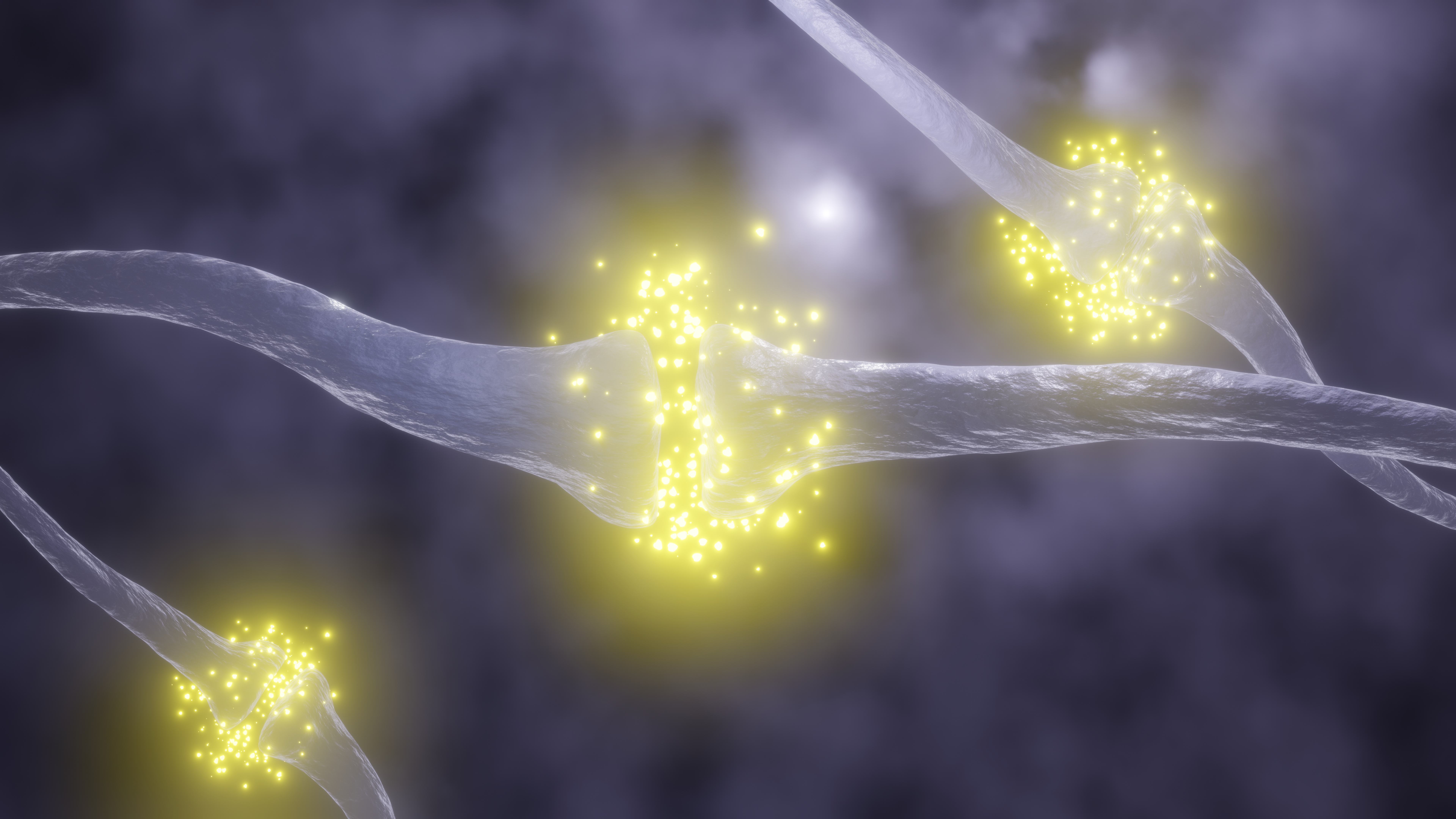
At the end of the axon lie the synaptic terminals, the small but critically important structures that connect the neuron to its targets. These endings are presented with clarity, shaped to emphasize their delicate form and their role in transmitting signals. The 3D environment shows the terminals positioned near surfaces that represent dendrites of other neurons, illustrating how communication occurs across tiny gaps through specialized pathways. The viewer can clearly understand that these regions serve as bridges for information transfer.
Surrounding the entire neuron, the digital landscape adds additional depth. Slight gradients, soft blurs, or faint textures hint at the environment within which neurons operate. This background does not distract from the main subject; instead, it provides a sense of space, making the illustration feel grounded within a larger biological setting. The result is an image that feels active, as if signals could appear at any moment along the pathways.
One of the greatest strengths of such a 3D rendering is how effectively it conveys the interconnected nature of neural networks. While only one neuron may be the primary focus, the subtle suggestions of nearby structures help viewers understand that this cell operates as part of an expansive communication system. Neurons depend on their connections to form pathways that handle tasks such as movement, memory, decisions, senses, emotions, and internal regulation. By showing these relationships visually, the illustration offers a powerful educational tool that communicates complexity without overwhelming the viewer.
This type of rendering also supports learning by simplifying the visualization of features that often appear abstract in textbooks. Instead of flat lines or symbols, the viewer sees a realistic three-dimensional model that demonstrates how each part relates spatially to others. The axon stretches ahead, the dendrites reach outward, and the terminals cluster at the edges. The entire structure becomes more understandable and memorable through this immersive perspective.
Beyond education, the illustration serves as a valuable tool for presentations, digital content, medical training, and scientific communication. Its clarity helps bridge the gap between scientific knowledge and public understanding, making neural structure more accessible to learners of all backgrounds. Researchers, teachers, and enthusiasts can use such visualizations to explain how the nervous system maintains coordination, awareness, balance, and control throughout the body.
In essence, a 3D rendered neuron illustration transforms a microscopic biological structure into a captivating visual experience. It captures the beauty of natural design, the complexity of communication pathways, and the importance of every structural element within the neuron. By presenting axons, dendrites, and synaptic pathways with exceptional clarity, the illustration deepens appreciation for the remarkable systems that sustain human life and cognition.